
Chapter 65: Disorders of the Bladder - PowerPoint PPT Presentation
1 / 80
Title:
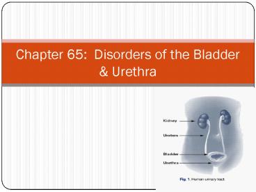
Chapter 65: Disorders of the Bladder
Description:
Signs of a bladder infection (eg, fever, chills, pain on urination) and ... The bladder can contract without warning, fail to accommodate adequate volumes ... – PowerPoint PPT presentation
Number of Views:199
Avg rating:3.0/5.0
Title: Chapter 65: Disorders of the Bladder
1
Chapter 65 Disorders of the Bladder Urethra
2
Overview Pg. 1153
- Disorders of the bladder urethra are common and
can be the source of severe problems that become
chronic, altering a clients lifestyle. - Many disorders affecting the bladder and urethra
are treated on an outpatient basis, but the more
serious disorders require hospitalization
3
Voiding Dysfunction
- Urinary retention urinary incontinence are
voiding dysfunctions - 1. Urinary retention is the inability to urinate
or effectively empty the bladder. - 2. Urinary incontinence is the inability to
control the voiding of urine. - Both require sensitivity to the clients needs,
both physiologic and psychosocial
4
Urinary Retention pg 1153
- May be either acute or chronic.
- Acute urinary retention is seen in complete
urethral obstruction, after general anesthesia,
or with the administration of certain drugs such
as Atropine or a Phenothiazine.
5
(No Transcript)
6
Urinary Retention
- Chronic urinary retention is often seen in
clients with disorders such as prostatic
enlargement or neurologic disorders that result
in a neurogenic bladder ( a bladder that does not
receive adequate nerve stimulation)
7
Urinary Retention
- The client with acute urinary retention usually
is not able to void at all. - The client with chronic urinary retention may be
able to void but does not completely empty the
bladder (retention with overflow) and has a large
residual volume. - The residual urine is urine retained in the
bladder after the client voids. The amount may
vary from 30 mL to several hundred milliliters.
8
S/S
- S/S Acute sudden inability to void, distended
bladder, and severe lower abdominal pain and
discomfort - S/S Chronic may produce no symptoms because the
bladder has stretched over time and accommodates
large volumes without producing discomfort.
9
S/S
- The overstretched bladder does not contract
effectively and the client is unaware that the
bladder is not emptying completely. - If the amount of residual urine is large, the
client may void frequently in small amounts. - Signs of a bladder infection (eg, fever, chills,
pain on urination) and dribbling of urine may
also be present.
10
(No Transcript)
11
Urinalysis
- How do you collect??????
- 24 hour Urine collection
- From indwelling foley catheter
- For pregnancy test
- For C S
12
Medical Management pg 1154
- Acute requires immediate catheterization.
- Chronic managed by permanent drainage with a
urethral catheter, suprapubic cystostomy tube (a
catheter inserted through the abdominal wall
directly into the bladder), or clean intermittent
catheterization (CIC)
13
(No Transcript)
14
Medical Management
- Permanent catheterization of the bladder carries
the risk of bladder stones, renal disease,
bladder infection, and urosepsis, a serious
systemic infection from microorganisms in the
urinary tract invading the bloodstream. - Clean intermittent catheterization (CIC) is the
preferred method.
15
Box 65-1 pg 1154
- Crede apply gentle downward pressure to the
bladder during voiding. This maneuver may be
done by the client or family member. The client
may also do this by sitting on the toilet and
rocking back and forth gently. - Valsalva instruct the client to bear down as
with defecation. Do not teach this method to a
client with cardiac problems or who may be
adversely affected by a vagal response (heart
rate slows)
16
Nursing Management
- An important nursing responsibility is measuring
I O, palpating the abdomen for a distended
bladder, promoting complete urination, and
monitoring the voiding pattern of clients
17
Acute Urinary Retention pg 1155
- Catheters are sized according to the French
system, for example, 14F to 24F. The higher the
number, the larger the diameter of the catheter.
- Examples of the various types of catheter tips
are shown in Figure 65-1
18
(No Transcript)
19
(No Transcript)
20
Acute Urinary Retention
- If the volume of urine is larger (gt 700 mL), it
may be necessary to clamp the catheter before the
bladder has emptied completely to prevent bladder
spasms or loss of bladder tone. - This practice varies so check agency policy
- NCLEX!!!!
21
Acute Urinary Retention
- Clients managed by CIC, establish a schedule.
They are catheterized every 4 to 6 hours
depending on the amount of urine obtained and the
fluid intake. - The bladder should not be allowed to get
distended beyond 350 mL because bladder over
distention results in loss of bladder tone,
decreased blood flow to the bladder, and
reduction in the layer of mucin that protects the
bladder mucosa.
22
Acute Urinary Retention
- CIC continues until the post void residual volume
is less than 30 mL - To obtain accurate results very important to
let the pt void first then immediately after the
attempt perform the catheterization. - Record both the volume voided (even if it is
zero) the volume obtained by catheterization. - Post-op urinary retention usually resolves within
24 to 48 hours.
23
Intermittent Catheterization pg 1155
- Hospital is sterile!!
- Home is clean rather than aseptic technique.
- They use a red rubber catheter that can be washed
and reused for 2 to 3 months before replacing. - Gloves are not required but client must wash
their hands thoroughly before and after the
procedure.
24
Intermittent Catheterization
- The schedule is usually 3 to 4 times a day,
although the frequency can be increased depending
on residual volume. - If more than 400 mL is returned, the client
should be catheterized more often.
25
Indwelling Catheters pg 1156
- A urethral indwelling catheter is one route for
permanent bladder catheterization - Cystostomy tube, also called a suprapubic
catheter, is an alternative that is inserted
through an abdominal incision into the bladder. - Clients require catheter care including careful
cleansing of the urethral meatus or cystostomy
site and proximal catheter, maintenance of the
integrity of the closed drainage system, proper
anchoring of the tube to avoid tension and
promote drainage.
26
(No Transcript)
27
Urinary Incontinence pg 1156
- May result either bladder or urethral dysfunction
(or both). - The bladder can contract without warning, fail to
accommodate adequate volumes of urine, or fail to
empty completely and become overstretched,
resulting in overflow incontinence. - These conditions result from neurologic disease,
prostatic enlargement, bladder outlet
obstruction, or trauma in all clients, and
bladder prolapse or low estrogen levels in women.
28
Urinary Incontinence
- A neurogenic bladder may be spastic, causing
incontinence, or it may be flaccid, causing
retention.
29
Incontinence
- Complain of urgency, frequency, leaking small
amounts when coughing or sneezing, or complete
inability to control urine, depending on the
underlying cause.Treatment medications, CIC,
surgeries - Nursing focuses on instruction on exercises to
increase muscle tone and voluntary control (Kegel
Exercises), techniques to assist bladder
emptying, and bladder training. 65-3 pg 1158
30
Cystitis pg 1160
- An inflammation of the urinary bladder.
- Usually caused by a bacterial infection.
- S/S urgency, frequency, low back pain, dysuria,
perineal and suprapubic pain, hematuria,
especially at the termination of the stream
(terminal hematuria) - If bacteria is present may have chills fever.
31
Medical Management
- Antimicrobial therapy and correction of
contributing factors. - Cranberry juice or vitamin C may be recommended
to keep the urine acidic and enhance the
effectiveness of drug therapy. - Cranberry juice helps to acidify the urine and
provides a less favorable climate for bacterial
growth. - Guidelines 65-4 Preventing cystitis
32
(No Transcript)
33
Interstitial Cystitis (IC) Pg. 1161
- A chronic inflammation of the bladder mucosa.
- The bladder wall contains multiple pinpoint
hemorrhagic areas that join and form larger
hemorrhagic areas that may progress to fissuring
and scarring of the bladder mucosa.
34
(No Transcript)
35
Interstitial Cystitis (IC)
- Superficial erosion of the bladder mucosa
(Hunners ulcer) may develop. - Eventually the bladder shrinks from scarring.
- S/S frequent, painful urination and passing a
small volume of urine are the most common
symptoms. - The pain may be described as searing or burning.
Has the need to void ASAP when a small amount of
urine is present in the bladder
36
(No Transcript)
37
Nursing Management
- Instruct to avoid spicy and acidic foods because
they may contribute to pain and discomfort. - Disrupts their daily lives due to the pain and
frequent trips to the bathroom to void. - Emotional support and referral to a chronic pain
center to cope and an support group.
38
Urethritis Pg. 1161
- Inflammation of the urethra (gt in men)
- Urethritis caused by microorganisms other than
gonorrhea is called nongonococcal urethritis. - May be secondary to vaginal infections.
- Soaps, bubble baths, sanitary napkins, or scented
toilet paper may also cause urethritis.
39
- MenChlamydia, irritation during vigorous
intercourse, rectal intercourse, or intercourse
with a woman who has a vaginal infection
40
S/S of Urethritis
- Discomfort on urination varying from a slight
tickling sensation to burning or severe
discomfort and urinary frequency. - Fever is not common, but fever in the male client
may be due to further extension of the infection
to areas such as the prostate, testes, and
epididymis.
41
Urethritis
- Tx antibiotic therapy, liberal fluid intake,
analgesics, warm sitz baths, good diet, rest. - !!!!!Even when a female patient has a foley
catheter, remember to clean from front to back.
!!!!!!
42
Obstructive Disorders Pg 1163
- Obstruction of the lower urinary tract is a
blockage in the bladder or in the urethra. - Many obstructions are r/t congenital anomalies,
but in adults, obstructions occur from stones
that block the passage of urine, or from a
narrowing that occurs as a result of a trauma,
inflammation, or infection.
43
Box 65-4 S/S
- Straining to empty bladder
- Feeling that bladder does not empty completely
- Hesitancy
- Weak stream
- Frequency
- Overflow incontinence
- Bladder distention
44
Bladder Stones pg 1163
- Large bladder stones develop in those with
chronic urinary retention and urinary stasis. - Clients who are immobile (eg, the unconscious
client or those with paraplegia or quadriplegia)
also may have a tendency to form bladder stones.
45
(No Transcript)
46
Assessment
- Symptoms of bladder stone formation include
hematuria, suprapubic pain, difficulty starting
the urinary stream, symptoms of a bladder
infection, and a feeling that the bladder is not
completely empty. - Some clients may have few or no symptoms.
47
Medical Management
- Bladder stones may be removed through the
transurethral route, using a stone-crushing
instrument (lithotrite). - This procedure, called a litholapaxy, is suitable
for small and soft stones and is performed under
general anesthesia. - Larger, non-crushable stones must be removed
through a surgical (suprapubic) incision into the
bladder.
48
(No Transcript)
49
Medical Management
- When it is possible to determine the chemical
composition of stones that have been passed or
removed, dietary treatment may be attempted to
adjust the pH of the urine to keep the urinary
salts in solution and thus prevent the formation
of stones.
50
(No Transcript)
51
Medical Management
- Uric acid stones may be prevented by a low-purine
diet - Increased fluid intake and the administration of
sodium bicarbonate may prevent the formation of
cystine stones. - Clients with a hx of calcium stone formation may
have to limit their intake of milk and milk
products.
52
Nursing Management
- Report any evidence of gross hematuria
IMMEDIATELY!! - Encourage fluids unless contraindicated by heart
failure or renal disease - Filter the urine for stones by straining all
urine through gauze or wire mesh. If solid
material is found, send it in a labeled container
to the lab for analysis.
53
(No Transcript)
54
Nursing Management
- For moderate to severe pain, administer a
narcotic analgesic as ordered - If undergoes a litholapaxy, a urethral catheter
may be left in place to keep the bladder
continuously empty for 1 to 2 days. - Monitor I O
- Encourage fluids once tolerated
55
Nursing Management
- If open removal is required, the bladder is
incised and the stone removed. - A urethral catheter may be left in place for a
week or more to keep the bladder empty and
prevent tension on the bladder sutures. - Instruct to contact the physician if hematuria,
burning, chills, fever, or pain occurs.
56
Urethral Strictures pg 1164
- Strictures of the urethra are caused by
infections such as untreated gonorrhea or chronic
nongonoccoccal urethritis. - Other causes include trauma to the lower urinary
tract or pelvis, such as accidents, childbirth,
intercourse, or surgical procedures. - May be congenital
- A stricture (narrowing) in the urethra obstructs
the flow of urine and can cause complications in
the bladder or upper urinary tract.
57
Urethral Strictures
- The kidney pelvis can become distended with the
backflow of urine. - The bladder distends when the urethra is
obstructed and a diverticulum (outpouching) of
the muscular bladder wall may form. (fig 65-5) - In some instances more than one diverticulum may
be seen
58
Urethral Strictures
- Urine becomes trapped in the diverticulum,
stagnates, and becomes a culture medium for
bacteria. - For this reason, infection occurs often and is
difficult to control until the obstruction is
corrected.
59
S/S
- Slow or decreased force of stream of urine,
hesitancy, burning, frequency,nocturia, and the
retention of residual urine in the bladder, which
may lead to bladder distention and infection. - The client may be able to pass more urine after
voiding and waiting a few minutes. - The final quantity of urine comes form the
diverticulum and may be malodorous.
60
Medical Management
- Treated by dilation, which is the use of
specially designed instruments called bougies,
sounds, filiforms, and followers. (fig 65-6) that
are passed gently into the urethra. - Although done gently, the procedure is usually
painful
61
(No Transcript)
62
- Because forceful stretching of the urethra may
cause bleeding and further stricture formation,
dilation begins with a 6F or 8F urethral dilator. - During subsequent treatments, the MD increases
the size of the dilator until a 24F or 26F can be
tolerated. - Periodic dilatations are usually required
indefinitely or until the condition is corrected
surgically.
63
(No Transcript)
64
Nursing Management
- Advise the client that the urine may be blood
tinged following urethral dilatation and that it
may burn when voiding. - Suggest sitz baths and nonnarcotic analgesics to
relieve discomfort. - Contact md if difficulty voiding or frank
bleeding occurs.
65
Malignant Tumors of the Bladder Pg. 1165
- Are frightening for clients
- Bloody urine is often the first sign of problems
and is the reason that clients seek medical
attention. - Malignant tumors of the bladder are the most
common tumors in the urinary system.
66
Hazards Include
- Exposure to industrial dyes, paint, or rubber
- Occupational exposure to sewage
- Coal gas
- Cigarette smoking and second-hand smoke
- Coffee drinking
- Use of artificial sweeteners
67
S/S
- First symptom of a malignant tumor of the bladder
is painless hematuria - UTI with symptoms such as fever, dysuria,
urgency, and frequency. - R/T Metastases pelvic pain, urinary retention,
and urinary frequency due to occupation of
bladder space by the tumor.
68
Medical Management
- Varies according to the grade and stage of the
tumor. - Resection of the tumors may be done. Have a high
incidence of recurrence, consequently, a
cystoscopic examination is performed every 2 to 3
months. - Clients having no recurrence of the tumor for at
least a year require cystoscopic examinations
every 6 months for the rest of their lives so
that recurrence of the tumor or a new malignant
growth can be detected early.
69
Surgical Management
- A cystectomy surgical removal of the bladder and
a urinary diversion procedure are necessary when
the tumor has penetrated the muscle wall. - When a cystectomy is performed, the bladder and
lower third of both ureters are removed.
70
Surgical Management
- Once a cystectomy is performed, urine must be
diverted to another collecting system. - This is called a urinary diversion
- Some urinary diversions require external ostomy
bags to collect the urine other types create a
reservoir within the body and the reservoir is
catheterized to drain the urine. - In some instances the urine is diverted to the
colon and the client voids rectally. - The more common types of urinary diversion
procedures are described in Table 65-7 pg 1167
71
Preoperative
- The client faces drastic changes in the manner of
excreting urine from the body, the diagnosis of
cancer, and the changes in body image - Encourage the client to talk about the surgery
and the changes that will occur. - Suggest a visit from a member of a local ostomy
group to provide emotional support as well as
information. - Photographs or drawings are useful in showing the
placement of the stoma and urostomy pouch.
72
Postoperative Period
- Management issues related specifically to urinary
diversion procedures include observing for
leakage of urine or stool from the anastomosis,
maintaining renal function, assessing for s/s of
peritonitis, maintaining integrity of the urinary
diversion and urine collection devices,
maintaining skin and stomal integrity,promoting a
positive body image, and teaching the client how
to manage the diversion.
73
Postoperative Period
- Label all urinary drainage tubes, and measure and
record the urine output from EACH catheter or
stoma every hour. - Record each measurement SEPARATELY!
- Maintaining accurate I O measurements during
the post-op period is important because it
indicates both renal function and the integrity
of the urinary diversion structures.
74
Postoperative Period
- Obstruction of urine flow can severely damage the
kidneys if urinary drainage stops or decreases
to less than 30 mL/hour, or if the client
complains of back pain, notify the physician
immediately. - Inspect the urine for color, clarity, and
presence of blood. - Immediately report concentrated, cloudy, or
bloody urine to the MD. - Ureteral stents will remain in place for several
days after surgery.
75
Postoperative Period
- Connect the NG tube to low intermittent suction.
This prevents distention and pressure on the
suture line due to the collection of gas in the
bowel. - The NG tube is removed once peristalsis has
returned and the diet can be advanced.
76
(No Transcript)
77
Trauma pg 1171
- Various types of injury can affect the urinary
tract. - Gunshot and stab wounds, crushing injuries,and
forceful blows can result in tears, hemorrhage,
or penetration of one or more parts of the
urinary tract. - Injuries to the kidney area may result in
bruising or tearing of the kidney and its
capsule. - Depending on the severity of the injury, blood
and urine may leak into the peritoneal cavity.
78
Assessment
- Symptoms vary according to the area affected and
the type of injury. - Anuria, hematuria, pain in the abdomen (which may
indicate bleeding or leakage of urine into the
abdominal cavity), pain in the bladder or kidney
areas, and symptoms of shock may be indicators of
urinary tract injury.
79
Assessment
- During treatment of a client with extensive
injury, an indwelling catheter may be inserted,
and hematuria or lack of urine output may be the
first sign that a traumatic injury to the urinary
tract injury has occurred. - Certain other types of injuries, such as stab or
gunshot wounds, may be immediately identified
because of outward signs of injury (eg, entry
wounds on the skin surface)
80
Management
- Tx depends on the type, location, and extent of
injury as well as on the condition of the client. - The most important nursing task is recognition of
abnormal findings. - Lack of urinary output, diffuse and severe
abdominal pain, and hematuria are examples of s/s
that may be indicative of an injury to the
urinary tract. - In some instances, the injury may be such that
symptoms DO NOT appear for several hours or days
after the initial trauma.
Recommended
CrystalGraphics Presentations


