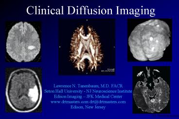
Clinical Diffusion Imaging - PowerPoint PPT Presentation
1 / 128
Title: Clinical Diffusion Imaging
1
Clinical Diffusion Imaging
- Lawrence N. Tanenbaum, M.D. FACR
- Seton Hall University - NJ Neuroscience Institute
- Edison Imaging JFK Medical Center
- www.drtmasters.com drt_at_drtmasters.com
- Edison, New Jersey
2
(No Transcript)
3
Diffusion imaging
- detection random motion of water molecules
- translation of water over short distances
- DW pulse sequence
- apply large gradient pulse
- impart phase modulation
- wait
- spins diffuse away randomly
- apply reverse pulse
- rephase (abnormal) stationary spins (retain
signal) - (normal) moving spins remain dephased
4
Diffusion Imaging
principles
Disease Causes Restricted Molecular Motion
Diffusion Gradients Sensitize to Motion
Increased Image Intensity
5
Diffusion imagingdefinitions
- individual direction images
- images obtained with diffusion gradient applied
in X, Y or Z direction - corpus callosum
- allows diffusion of water molecules along
orientation of fibers - restricts diffusion of water perpendicular to
fiber orientation
6
Diffusion imagingisotropic diffusion image
- isotropic images
- combined, trace weighted images
- combination of individual direction images
7
Diffusion imagingisotropic diffusion image
- advantages
- direction specific variations in diffusion
eliminated - increase conspicuity of subtle ischemia
- improved SNR
8
TechniqueDW-EPI
- 8200 / min
- B 800 (ES, HS) 1200 (TS) s / mm2
- single shot SE EPI
- 5 mm / 0 mm
- 96 x 128, 1 nex
- 24 x 19 FOV
- 30 locations, 3 directions, 033
- trace weighted, B0 images generated by scanner
Mardi Gras NO 2001
9
1000
B value
10
TwinSpeed
B 1200 TE 69.9
B 2000 TE 79.1
B 3000 TE 87.6
B 4000 TE 94.4
B 6000 TE 105.2
B 5000 TE 100.2
B 7000 TE 109.7
11
high SNR
DWI
12
no ASSET
8 channel head coil 1.5 T
ASSET
13
1200 B 3 Tesla
ASSET
no ASSET
14
153
253
30 slc
15
no ASSET
ASSET
16
ASSET x 2
17
PROPELLER FSE Periodically Rotated Overlapping
ParallEL lines with Enhanced Reconstruction
18
(No Transcript)
19
DWI
Propeller FSE
ssEPI
20
3T Diffusion imaging
Propeller FSE
ssEPI
21
(No Transcript)
22
(No Transcript)
23
propeller
EPI
24
Conventional DWI PROPELLER DWI
Images courtesy of Barrow Neurological Institute
25
(No Transcript)
26
Diffusion imagingindications
- TIA and infarction
- demyelinating disease
- lesion characterization
- tractography
Steamboat 2001
27
Diffusion imaging
- conventional MRI techniques limited in the
detection of hyperacute stroke - DW techniques sensitive within minutes of onset
of infarction
28
Diffusion imaginginfarction
- diffusion measurably slower in regions of
ischemia when compared to normal brain - cell swelling with restriction of motion between
cells - intracellular shift of water (cytotoxic edema)
- slower diffusion environment
29
Diffusion imagingprinciples
- diffusion gradients sensitize MR Image to motion
of extracellular water - more motion darker image
Freely Diffusing Water Dark
Restricted Diffusion Bright
30
(No Transcript)
31
(No Transcript)
32
(No Transcript)
33
B 1200
34
(No Transcript)
35
(No Transcript)
36
(No Transcript)
37
(No Transcript)
38
(No Transcript)
39
Hyperacute infarction
35 minutes
40
Hyperacute infarction
35 minutes
41
(No Transcript)
42
1.5 T
0.2 T
43
(No Transcript)
44
Line Scan DWI
45
0.2T
46
Technique0.2 T Profile LSDI
- SR 25 gradients
- 367 / min
- B 800 s / mm2 , S-I direction
- 7.5 mm / 0 mm
- 64 x 64, 1 nex, 2.02 kHz BW
- 26 FOV, fAP
- 18 locations, 741
Orlando 2000
47
0.35T LSDI
48
0.35T ssEPI
49
0.35 T
DW EPI b value 1000 s/mm2 128 x 128 whole
brain in 20 seconds
50
(No Transcript)
51
ssEPI
0.7T OpenSpeed
LSDI
52
(No Transcript)
53
(No Transcript)
54
(No Transcript)
55
(No Transcript)
56
Open MR Diffusion Imaging Comparison of Line
Scan Diffusion Imaging at 0.2T With Diffusion
Weighted EPI at 1.5T in Patients With Recent
Infarction
- Tanenbaum LN, Borden N, Eshkar NE, Verro P, Sen S
- JFK Medical Center-New Jersey Neuroscience
Institute - Seton Hall School of Graduate Medical Education
- Edison, New Jersey
57
Materials and Methods
- 21 consecutive patients with symptoms of recent
infarction and positive findings on 1.5T DWEPI
(GE Signa SR120) were evaluated on a clinical
0.2T open system (GE Profile SR17) with LSDI.
58
Overall comparisonFriedman test
- 0.2T LSDI (plt0.0001) and DWEPI 1.5T (plt0.0001)
superior to 1.5 T FLAIR in the detection of
recent infarction
59
ResultsSensitivity 0.2T LSDI
- 22 recent infarcts seen on DWEPI at 1.5T
- Readers 1 and 2
- 20 definite infarcts (90.9)
- 1 possible infarct (4.5 )
- 1 no infarction (4.5 )
60
(No Transcript)
61
(No Transcript)
62
(No Transcript)
63
(No Transcript)
64
(No Transcript)
65
(No Transcript)
66
(No Transcript)
67
(No Transcript)
68
1.5T FLAIR
1.5T DWEPI
0.2T DWLSI
69
Diffusion imagingdefinitions
- diffusion weighted image
- randomly moving spins dephase
- see relatively LOW signal in normal tissues
- relatively static spins rephase
- see HIGH signal in areas of low diffusion
- ADC map
- isotropic image of apparent diffusion
coefficient, trace image - areas of low diffusion have LOW signal intensity
70
e-b(ADC)
T2W e-b(ADC)
Trace weighted
ADC
Exponential ADC
71
(No Transcript)
72
(No Transcript)
73
(No Transcript)
74
(No Transcript)
75
(No Transcript)
76
(No Transcript)
77
(No Transcript)
78
(No Transcript)
79
Diffusion imaginginfarction
- ADC decreases 30 - 60 with ischemia in
experimental models. - with permanent vascular occlusion ADC decreases
highly correlate with areas of ultimate
infarction volume. - regional decreases in ADC can reverse after early
reperfusion - reversal accurately predicts size of infarction
80
Lesion aging
T2
FLAIR
DW-EPI
Tampa 2001
81
1.5 T
0.2 T
Steamboat 2001
82
(No Transcript)
83
(No Transcript)
84
0.2T DWI
85
Evaluation of Diffusion Weighted EPI and FLAIR
in Patients with Subacute Ischemic Symptoms
- LN Tanenbaum, BA Johnson, BP Drayer,
- JC Friel, P Verro and RG Hoffman
- New Jersey Neuroscience Institute
- Seton Hall School of Graduate Medical Education
- Edison, New Jersey
86
Results Change in diagnosis with DW-EPI29
patients with clinical stroke
- no infarct
- possible infarct to additional infarct 4
(7) - definite infarct
- no infarct
- possible infarct to definite infarct 35
(59) - possible infarct
- definite infarct to no infarct
5 (8) - no change from FLAIR 15
(25)
87
(No Transcript)
88
(No Transcript)
89
(No Transcript)
90
venous infarction
91
Diffusion imaginginfarction
- sensitive almost immediately
- appears to reflect clinical outcome
- distinguish between TIA and silent CVA
- differentiate acute and chronic infarction
- socioeconomic impact
Pusan 2000
92
Lesion characterization
new onset seizures
93
(No Transcript)
94
Case study Diagnosis Infarction
95
Lesion characterization
96
Lesion characterization
ADC map
97
vasogenic edema
BBBB
DWI
ADC
98
Diffusion imagingdemyelinating disease
- characterize and detect regions of active
demyelination - most / many contrast enhancing lesions show high
signal on (moderate B) DW images - some lesions with altered diffusion do not
enhance at 0.1 mmol/kg - enhance at 0.2 - 0.3 ?
- avoid (cost of) gadolinium?
Kuala Lumpur 2000
99
(No Transcript)
100
(No Transcript)
101
(No Transcript)
102
(No Transcript)
103
(No Transcript)
104
(No Transcript)
105
(No Transcript)
106
(No Transcript)
107
(No Transcript)
108
(No Transcript)
109
(No Transcript)
110
(No Transcript)
111
(No Transcript)
112
1.5 T Excite 8 channel coil 1024 x 384 3 mm
113
(No Transcript)
114
(No Transcript)
115
Trace
B0
ADC
Exponential
116
(No Transcript)
117
(No Transcript)
118
(No Transcript)
119
(No Transcript)
120
(No Transcript)
121
(No Transcript)
122
(No Transcript)
123
(No Transcript)
124
(No Transcript)
125
(No Transcript)
126
(No Transcript)
127
(No Transcript)
128
(No Transcript)
129
(No Transcript)
130
(No Transcript)
131
(No Transcript)
132
(No Transcript)
133
hematoma
134
(No Transcript)
135
(No Transcript)
136
(No Transcript)
137
prolonged seizure activity
138
lymphoma
M. Maya MD
139
lymphoma
M. Maya MD
140
www.drtmasters.com

