
Catalytic Mechanism and Strategies - PowerPoint PPT Presentation
1 / 43
Title:
Catalytic Mechanism and Strategies
Description:
... Synthase - a simple energy producing system. H H H H ADP ... between the Schiff base. and Asp-85 'Switch' and proton. release to the extra- cellular surface ... – PowerPoint PPT presentation
Number of Views:83
Avg rating:3.0/5.0
Title: Catalytic Mechanism and Strategies
1
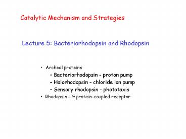
Catalytic Mechanism and Strategies
- Lecture 5 Bacteriorhodopsin and Rhodopsin
- Archeal proteins
- Bacteriorhodopsin - proton pump
- Halorhodopsin - chloride ion pump
- Sensory rhodopsin - phototaxis
- Rhodopsin - G protein-coupled receptor
2
bR and ATP Synthase - a simple energy producing
system
H
light
H
H
H
H
H
bacteriorhodopsin
ATP synthase
ADP
ATP
Proton gradient created by bR is used to drive
ATP synthesis
3
Photoreaction cycle of retinal chromophore
N-Lys
bR568
H
H
N
H
K640
Lys
N
M412
H
Lys
4
Bacteriorhodopsin Photocycle
5
Isomerization of the retinal to 13-cis
6
Protonation equilibrium between the Schiff base
and Asp-85
7
Switch and proton release to the
extra- cellular surface
8
Reprotonation of the Schiff base by Asp-96
9
Reprotonation of Asp-96, reisomerization of
the retinal to all-trans
10
Deprotonation of Asp-85
11
Conformational changes related to
function what causes steps 1 - 5?
a
12
Photoreaction cycle of retinal chromophore
Asp 96 - COOH
Proton donor
in
direction of proton pumping
N
bR568
H
W402
out
Asp 85 - COO-
W401
Proton acceptor
W406
Arg 82 - NH2
W402 water 402
W403
W404
Hydrogen-bonded network with well-defined structur
al water molecules on extracellular side of the
protein Initial direction of proton motion is
opposite to the direction of proton pumping!
Glu 194, 204
H
13
Photoreaction cycle of retinal chromophore
in
direction of proton pumping
N
bR568
H
out
Asp 96 - COOH (high pKa)
H
pKa of Schiff base drops
N
NH N H Ka HN/NH
K640, L550
Asp 85 - COO-
Arg 82 - NH2
pKa of Asp85 increases
14
H2O
Photoreaction cycle
Asp 96 - COOH
in
direction of proton pumping
N
M412
out
Asp 85 - COOH
W401
W406
Arg 82 - NH2
Glu-C00- HOOC-Glu
Proton transfers to Asp85 Arg 82 swings down
toward Glu194/Glu204 Glu204 deprotonates Helix 6
moves and allows water exposure to Asp96
H
15
H2O
Photoreaction cycle
Asp 96 - COO-
in
direction of proton pumping
H
N
N520
out
Asp 85 - COOH
W401
W406
Arg 82 - NH2
-OOC-Glu
Asp96 deprotonates Proton transferred to Schiff
base Retinal reisomerizes
H
16
H
H2O
Photoreaction cycle
Asp 96 - COOH
in
direction of proton pumping
O640
N
H
out
Asp 85 - COOH
W401
W406
Arg 82 - NH2
Retinal reisomerizes Helix 6 moves back and
forces water out Asp96 reprotonates Asp85
deprotonates (due to return of NH) Glu204
reprotonates, Arg82 swings back toward Asp85
-OOC-Glu
17
1.55 Å crystal structure reveals functional
water molecules
18
The BR state
Hydrogen-bonds at active center
19
Hydrogen-bonded network around water 402
20
The BR state
Hydrogen-bonds at active center
A hydrogen-bonded network links Asp-85 to
Glu-194/Glu-204
21
The BR state
A chain of covalent and hydrogen-bonds links
region of retinal to Asp-96
Hydrogen-bonds at active center
A hydrogen-bonded network links Asp-85 to
Glu-194/Glu-204
22
H-bond network on intracellular side is not
continuous
23
(No Transcript)
24
BR
25
K
26
M1
27
M2
28
M2
29
G protein-coupled receptors are involved in many
signal transduction cascades and are a major
targetof pharmaceuticals
- Visual receptors rhodopsin, cone
pigments - Olfactory receptors
- Hormone receptors follicle stimulating
hormone thyrotropin
receptor - Neurotransmitters dopamine receptor
- Metabotropic receptors glutamate receptor
30
G protein-coupled receptors
Ligand
- 7 transmembrane helices
- Ligands bind from the extracellular side
- Heterotrimeric G protein binds from the
cytoplasmic side - Conserved residues in transmembrane helices
b
GDP
a
g
31
G Protein-Coupled Receptors
Light
Ligand
- Receptor conformational change
- upon ligand binding.
- Binding of heterotrimeric G protein.
- Exchange of GDP for GTP in a-subunit.
b
b
GDP
g
a
g
GTP
a
32
Light
Each disc membrane contains many photoreceptors
Rod Cell
Outer segment contains about 2000 disc membrane
Each photoreceptor is a protein containing a
small molecule, vitamin A
Nerve impulse
33
Color Vision
- 11-cis retinal is
- the same in all
- visual pigments
- Without bound
- retinal the protein
- is colorless.
- Interaction
- between
- the retinal and
- protein changes
- the color of the
- retinal.
11-cis retinal lmax 380 nm
green cone protein
green cone pigment
11-cis retinal lmax 380 nm
blue cone protein
blue cone pigment
11-cis retinal lmax 380 nm
red cone pigment
red cone protein
34
Vitamin A (retinal) is the photoreactive molecule
in photoreceptors
Light
Cis, bent
N-Lys 296
11-cis retinal (inactive)
H
Trans, straight
all-trans retinal (active)
35
Energy storage in rhodopsin
Light
hydrophobic
N
H
Glu113 - COO-
hydrophilic
H
N
36
Energy storage in rhodopsin
H
N
N
Distance 5.0 Å
H
Distance 2.5 Å
Glu113 - COO-
W k q1 q2/er -66 kcal/ mol ( r 2.5 Å, e
2)
W k q1 q2/er -33 kcal/ mol ( r 5.0 Å, e
2)
Energy stored 33 kcal/mol
37
Activation of rhodopsin
?G 40 kcal/mol
?Gstored 33 kcal/mol
11-cis-retinal
All-trans-retinal
38
Activation of rhodopsin
?-electrons go into anti-bonding orbitals CC
effectively becomes C-C
No barrier to isomerization - isomerization
takes 200 fs !
Isomerization occurs in the excited electronic
state
11-cis-retinal
H
All-trans-retinal
N-Lys
39
Helix interactions in GPCRs are critical.
?
GDP
?
?
Inactive
Active
40
Structural model of rhodopsin
IV
II
III
Glu113
-
H
I
N
V
VII
VI
Schiff base and Glu113 form a stable salt bridge
41
Structural model of rhodopsin
IV
II
III
Glu113
-
Gly121
H
I
N
V
Trp265
Tyr268
VII
VI
Shape and charge of retinal PSB is complementary
to protein binding site.
42
Structural model of rhodopsin
IV
II
III
Glu122
Glu113
-
His211
H
Arg135
I
N
V
Glu247
VII
VI
Helix interactions lock the receptor in the
off-state in the dark.
43
Isomerization of retinal breaks helix interactions
IV
II
III
Glu113
H
I
V
N
VII
VI
Recommended
CrystalGraphics Presentations


