
EEG - PowerPoint PPT Presentation
1 / 69
Title: EEG
1
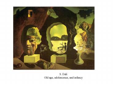
S. Dali Old age, adolescence, and infancy
2
Brain Imaging
- Ben H. Jansen
- ECE Dept, U-Houston
17th century BC Smith Papyrus
ys
3
Brain Imaging
- Anatomy
- Photography
- Light, x-ray
- Tomography
- CAT
- MRI
- Activity
- Electrical
- Magnetic
- Function
- fMRI
- PET
- SPECT
CAT
PET
Photography
MRI
4
Brain
5
Neurons
6
Columns and Layers
7
ElectroEncephaloGram Discovery
Ancient Greeks Had no word for brain. Head
Kephale. Supposed that the mind was located in
the midriff (diaphragm, phren). Schizophrenia.
Galvani (1790) showed that (dead) muscle tissue
can contract when electrically stimulated
Fritsch and Hitzig (1870) Medical officers of
the Prussian army. Applied electrical stimulation
to the exposed brain of victims in the Sedan war.
Noticed a contraction of the right aide of the
body if a stimulus was applied to the left
hemisphere.
8
Facsimile of the Surrender Note sent by Napoleon
III. to King Wilhelm I. of Prussia at Sedan, 1st
September 1870
Nayant pas pu mourir au milieu de mes troupes,
il ne me reste qua remettre mon épée entre les
mains de Votre Majesté. Je suis de Votre Majesté
le bon frère. Napoleon
9
EEG Discovery
Caton (1875) discovered that the brain produced
electricity.
Prawdwicz and Neminsky (1913) obtained
electrocerebrogram from dog using galvanometer
without amplifier.
10
Hans Berger, Jena, Germany, 1929
Berger used galvanometer which caused deflections
of a light beam, which were photographed.
Zinc plate electrodes stuck in epidural tissue of
patients with part of their skull removed.
11
Electroencephalogram Source
EEG corresponds to EPSPs (A-C) and IPSPs (B-D)
and not to APs
12
EEG Measurement
13
EEG Electrode locations
14
Electro-Corticogram
15
EEG Spontaneous activity
Generally less than 300 mV
Gamma 30-80 Hz
16
EEG from Age 1 Month to Adulthood
17
EEG Spontaneous Activity
18
Normal Patterns of Wakefulness in Adults
19
Light Sleep (adult)
20
Deep Sleep (adult)
21
Somnogram
22
Jane AntoniSlumber1993Dakis Jammon Collection,
Athens
23
Eye Movement Artifacts
24
Extracerebral Artifacts
25
Non-biological Artifacts
26
Photic Driving
27
Tonic-Clonic Seizure
28
Tonic-Clonic ltcontgt
29
Tonic-Clonic ltc-contgt
30
Evoked Potentials Characteristics
- Positive and/or Negative components at specific
latencies - Exogenous
- Putative
- Early/mid-latency (lt40ms)
- Related to stimulus characteristics
- Endogenous
- Cognitive processing
- Latency gt250ms
31
Ensemble Averaging
AEP 1 kHz, 50 ms tone to left ear Vertex to
right mastoid negative is up.
Ensemble average
Plus/minus average
32
Visual Evoked Potential Checkerboard Pattern
33
VEP Checkerboard Pattern
34
VEP Pattern Reversal
35
Somato-Sensory EP
36
Auditory Evoked Potentials
37
AEP
38
Auditory EPs
39
AEP and Stimulus Intensity
40
P300 and the Odd-Ball Paradigm
2 sec
F
F
F
F
F
F
R
R
R
F
F? Frequent/Non-Target stimulus
P300 response to a relevant, infrequently
occurring stimulus.
R? Rare/Target stimulus
41
Event Related Potential P300
Frontal
Occasional tones
Vertex
Frequent tones
Parietal
Reaction time
42
Contingent Negative Variation
Negative potential following a warning stimulus
and preceding a second stimulus requiring a
response.
43
Readiness (Bereitschafts) Potential
44
Magnetoencephalography - Definition
- Non-invasive brain imaging technique
- Passive measurement of minute current dipoles and
corresponding magnetic moments - Magnetic field generated by neurons on the order
of tens of femto (10-15)Tesla (the background
magnetic field of the earth is roughly 60 micro
(10-6)Tesla - High resolution in both space (2 - 3mm) and time
(lt1ms)
45
MEG - Apparatus
- Screen is used for patient stimulation for
functional mapping - Patient wears a helmet containing an array of
100 sensitive magnetic field measurement devices - Measurement devices are called SQUIDs
Superconducting Quantum Interference Devices
(kept at 7 degree Kelvin) - Measurements must occur in costly magnetically
shielded room
Clinical System by VSM Medtech
46
Measuring Functional Activity
- Its all in the blood
47
Tomography/Backprojection
48
Coronal Slice, Pathology
49
What Does (f)MRI Measure?
- Big magnetic field
- protons (hydrogen molecules) in body become
aligned to field - RF (radio frequency) coil
- radio frequency pulse knocks protons over
- as protons realign with field, they emit energy
that coil receives (like an antenna) - Gradient coils
- make it possible to encode spatial information
- MR signal differs depending on
- concentration of hydrogen in an area (anatomical
MRI) - amount of oxy- vs. deoxyhemoglobin in an area
(functional MRI)
50
MRI vs. fMRI
MRI
high resolution (1 mm)
fMRI
low resolution (3 mm but can be better)
one image
fMRI Blood Oxygenation Level Dependent (BOLD)
signal indirect measure of neural activity
many images (e.g., every 2 sec for 5 mins)
? neural activity ? ? blood oxygen ? ?
fMRI signal
51
BOLD signal
Blood Oxygen Level Dependent signal
- neural activity ? ? blood flow ? ? oxyhemoglobin
? ? T2 ? ? MR signal
Source fMRIB Brief Introduction to fMRI
52
fMRI Activation
Flickering Checkerboard OFF (60 s) - ON (60 s)
-OFF (60 s) - ON (60 s) - OFF (60 s)
Brain Activity
Source Kwong et al., 1992
Time ?
53
fMRI Activation
Source Posner Raichle, Images of Mind
54
Activation Statistics
Functional images
Time
Source Jody Culhams fMRI for Dummies web site
55
fMRI Equipment
Gradient Coil
Magnet (4T)
4T magnet
RF Coil
gradient coil (inside)
Head Coil
Surface Coil
Source Joe Gati, photos
Source Jody Culhams fMRI for Dummies web site
56
fMRI Setup
57
Magnet Safety
The whopping strength of the magnet makes safety
essential. Things fly Even big things!
Source www.howstuffworks.com
Source http//www.simplyphysics.com/ flying_objec
ts.html
58
PET/SPECT
- PET measures emissions from radioactively
labeled chemicals that have been injected into
the bloodstream - cyclotron to "label" specific drugs or analogues
of natural body compounds, such as glucose, with
small amounts of radioactivity. - Using different compounds, PET can show blood
flow, oxygen and glucose metabolism, and drug
concentrations in the tissues of the working
brain. - Single Photon-Emission Tomography (SPECT)
- uses isotopes with longer half-lives that can be
stored on site. - lower resolution (9x9x9 mm.)
- used to study the neurotransmitter dopamine.
59
Positron Emission Tomography
- A positron is an anti-electron.
- Positrons are given off during the decay of the
nuclei of specific radioisotopes. - When a positron meets an electron, the collision
produces two gamma rays - The gamma rays leave the patients body and are
detected by the PET scanner.
60
Isotopes
61
PET Scanner
62
Optical Topography
63
Intrinsic Signal
Source of Intrinsic Signals
Light reflectance of living tissue is activity
dependent
Major Source
Metabolic Activity
Rat brain and kidney tissue Chance et al.,
Science (1962) 137, 499
- Blood oxygenation HbO2?Hbr
- Blood volume change
Electrical Activity
Shore crab nerve trunk of the walking leg Hill
et al., J Physiol (1949) 108, 278
Light scattering component
64
Metabolic Intrinsic Signal
Metabolic Intrinsic Signal
Neurons
Capillaries
HbO2
Hbr
Oxygen
50m
50m
Harrison et al, Cereb Cortex (2002) 12, 225
Ekstrand et al, J Comp Neurol (2001) 434, 308
65
Cat Visual Cortex
Cat Visual Cortex
Orientation Selectivity
Hubel Wiesel, J. Physiol. 1959
66
OIIS
Optical Imaging of Intrinsic Signal
Monkey cat visual cortex Orientation preference
Blasdel Salama, Nature 1986
Grinvald et al., Nature 1986 Bonhoeffer
Grinvald, Nature 1991
Grinvald Bonhoeffer, in Brain Imaging 1996
67
(No Transcript)
68
Vincent van Gogh
69
(No Transcript)
Recommended
CrystalGraphics Presentations


