
A FACILITY FOR STUDYING BLOOD FLOW SHEAR STRESSES AND ENDOTHELIAL CELL ADHESION - PowerPoint PPT Presentation
1 / 2
Title:
A FACILITY FOR STUDYING BLOOD FLOW SHEAR STRESSES AND ENDOTHELIAL CELL ADHESION
Description:
A new CFI / OIT / UWO funded flow facility will be used to quantify this ... regions of laminar or turbulent pulsatile flow that model the cardiac cycle. ... – PowerPoint PPT presentation
Number of Views:46
Avg rating:3.0/5.0
Title: A FACILITY FOR STUDYING BLOOD FLOW SHEAR STRESSES AND ENDOTHELIAL CELL ADHESION
1
A FACILITY FOR STUDYING BLOOD FLOW SHEAR STRESSES
AND ENDOTHELIAL CELL ADHESION
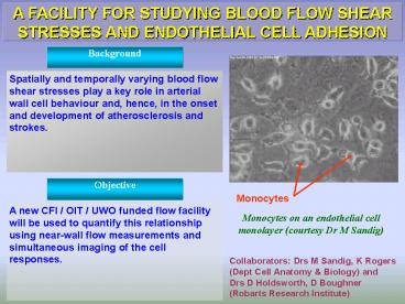
Background
Spatially and temporally varying blood flow shear
stresses play a key role in arterial wall cell
behaviour and, hence, in the onset and
development of atherosclerosis and strokes.
Objective
Monocytes
A new CFI / OIT / UWO funded flow facility will
be used to quantify this relationship using
near-wall flow measurements and simultaneous
imaging of the cell responses.
Monocytes on an endothelial cell monolayer
(courtesy Dr M Sandig)
Collaborators Drs M Sandig, K Rogers (Dept Cell
Anatomy Biology) and Drs D Holdsworth, D
Boughner (Robarts Research Institute)
2
Research To Be Carried Out
The facility, shown here, gives regions of
laminar or turbulent pulsatile flow that model
the cardiac cycle. Non-intrusive, micro-scale
measurements of the flow and shear stresses will
be made using optical anemometry (LDV), whilst
the response of live cultured endothelial cells
(on a coverslip) to this forcing cycle will be
determined using Laser Scanning Confocal
Microscopy (LSCM).
Expected Outcomes
A full description of the role of hemodynamic
forces in the different stages of the onset and
development of stenoses to give a better
understanding of why these diseases develop in
the way they do.
Recommended
CrystalGraphics Presentations


