
Sensory Systems - PowerPoint PPT Presentation
1 / 49
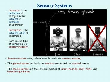
Title: Sensory Systems
1
Sensory Systems
- Sensation is the detection of changes in the
internal or external environment - Perception is the interpretation of sensations
- Each unique type of sensation is a sensory
modality
- Sensory neurons carry information for only one
sensory modality - The general senses are both the somatic senses
and the visceral senses - The special senses are the sense modalities of
vision, hearing, smell, taste, and
balance/equilibrium
2
Sensory Receptors
- Sensation usually involves 4 events
- Stimulation of a sensory receptor
- Transduction of the stimulus
- Generation of nerve impulses
- Integration of sensory input
- Sensory receptors can be classified by structure
- Free nerve endings are bare dendrites, which
produce generator potentials - Encapsulated nerve endings are enclosed in
special structures - Special receptor cells synapse with sensory
neurons to produce receptor potentials
3
The Five Senses
Vision
Hearing
Taste
Smell
Touch
Temperature Pain Balance Stretch Acceleration Pres
sure Texture Vibration Tickle Itch Etc.
4
Sensory Receptors Sensory Modalities
5
Sensory Receptors
6
Sensory Receptors
7
Sensory Receptors
8
Sensory Receptors
9
Sensory Receptors
10
Sensory Receptors
11
Temperature
- Thermoreceptors are free nerve endings
- There are two basic thermal sensations cold and
warm - Cold receptors mostly synapse with large
myelinated A fibers - Warm receptors mostly synapse with small
unmyelinated C fibers - Hot sensations activate both warm and cold
receptors, and pain receptors
12
Pain
- Nociceptors are free nerve endings
- There are two basic types of pain
- Fast pain is carried by large myelinated A fibers
- Fast pain is sometimes known as sharp, piercing,
pricking, emergency pain - An example of fast pain would be a knife cut or
stab wound - Slow pain is carried by small unmyelinated C
fibers - Slow pain is sometimes known as dull, aching,
throbbing, burning, or reminding pain - An example of slow pain would be a toothache, or
the day after an ankle sprain
13
Olfactory Epithelium
- 1 square inch of membrane holding 10-100 million
receptors - Covers superior nasal cavity and cribriform plate
- Odorants bind to receptors
- Na channels open
- Depolarization occurs
- Nerve impulse is triggered
14
Adaptation Odor Thresholds
- Adaptation decreasing sensitivity
- Olfactory adaptation is rapid
- 50 in 1 second
- Complete in 1 minute
- Low threshold
- Only a few molecules need to be present
- Methyl mercaptan added to natural gas as warning
- Hyposmia decreasing ability to smell
- 50 over age 65
- 75 over age 80
- Cigarette smoking
- Anosmia cannot smell
15
Gustatory Sensation Taste
- Taste requires dissolving of substances
- Five classes of stimuli sour, bitter, sweet,
salty, umami - Other tastes are a combination of the five
taste sensations plus olfaction - There may be a sixth taste bud, perhaps fatty
- Vallate papillae contain 100 - 300 taste buds
- Fungiform papillae contain 5 taste buds each
- Filiform papillae contain tactile receptors but
no taste buds - 10,000 taste buds found on tongue, soft palate
larynx
16
Physiology of Taste
- Receptor potentials developed in gustatory hairs
cause the release of neurotransmitter that gives
rise to nerve impulses - Complete adaptation in 1 to 5 minutes
- Thresholds for tastes vary among the 5 primary
tastes - Most sensitive to bitter (poisons)
- Least sensitive to salty and sweet
17
Vision
- More than half the sensory receptors in the human
body are located in the eyes - A large part of the cerebral cortex is devoted to
processing visual information - Eyeball is about 1 inch in diameter
- Over 80 of the eyeball is enclosed in the orbit
18
Lacrimal Apparatus
- About 1 ml of tears produced per day
- Spread over eye by blinking
- Contains bactericidal enzyme lysozyme
19
Tunics (Layers) of Eyeball
- The eye is constructed of three layers
- Fibrous Tunic(outer layer) sclera cornea
- Vascular Tunic (middle layer) choroid,
ciliary body, iris, lens - Nervous Tunic(inner layer) retina
20
Cavities of the Interior of Eyeball
- Anterior cavity (anterior to lens)
- Filled with aqueous humor
- Produced by ciliary body
- Continually drained
- Replaced every 90 minutes
- Two chambers
- Anterior chamber between cornea and iris
- Posterior chamber between iris and lens
- Posterior cavity (posterior to lens)
- Vitreous chamber filled with vitreous body
(jellylike) - Formed once during embryonic life
- Floaters are debris in vitreous of older
individuals
21
Muscles of the Iris
- Constrictor pupillae (circular) are innervated by
parasympathetic fibers while Dilator pupillae
(radial) are innervated by sympathetic fibers - Response varies with different levels of light
22
Electromagnetic Spectrum
Its not exactly correct, but you can get a rough
approximation by thinking of violet as 400 nm,
green as 500 nm, yellow as 600 nm, and red as 700
nm.
23
Photoreceptors Rods Cones
- Rods rod shaped
- Shades of gray in dim light
- 120 million rod cells
- Shapes movements
- Distributed along periphery
- Cones cone shaped
- Sharp, color vision
- 6-8 million
- Fovea of macula lutea
- Densely packed region
- At exact visual axis of eye
- Sharpest resolution (acuity)
24
Pathway of Nerve Signal in Retina
- Light penetrates retina
- Rods cones transduce light into action
potentials - Rods cones excite bipolar cells
- Bipolars excite ganglion cells
- Axons of ganglion cells form optic nerve leaving
the eyeball (blind spot) - To thalamus then the primary visual cortex
25
Visual Processing in Retina
- The layers of cells (bipolar, amacrine,
horizontal, ganglion) in the retinal process
information before it leaves the eye - For example, the presence of center-on and
center-off fields enhances contrast
26
Pathway of Nerve Signal to the Brain
- Medial optic fibers cross at the optic chiasm
- Lateral optic fibers remain on the same side of
the brain - Thus both sides of the brain get information from
both eyes
27
Refraction by the Cornea Lens
- Image focused on retina is inverted reversed
from left to right - Brain learns to work with that information
- 75 of refraction is done by cornea, the rest is
done by the lens - Light rays from gt 20 are nearly parallel and
only need to be bent enough to focus on retina - Light rays from lt 6 are more divergent need
more refraction - extra process needed to get additional bending of
light is called accommodation
28
Correction for Refraction Problems
- Emmetropic eye (normal)
- Can refract light from 20 ft away
- Myopia (nearsighted)
- Eyeball is too long from front to back
- Glasses concave
- Hypermetropic (farsighted)
- Eyeball is too short
- Glasses convex (coke-bottle)
- Astigmatism
- Corneal surface wavy
- Parts of image out of focus
29
Physiology of Vision
- Photopigments undergo structural changes upon
light absorption - Retinal is the light absorbing part of all visual
photopigments - All photopigments involved in vision contain a
glycoprotein called opsin and a derivative of
vitamin A called retinal
- There are four different opsins
- A cone contains one of three different kinds of
photopigments so there are three types of cones - Permit the absorption of 3 different wavelengths
(colors) of light - Rods contain a single type of photopigment
(rhodopsin)
30
Cone Absorption Spectra
31
Color Blindness
- Color blind individuals are missing one or more
types of opsin - As a result they may see some colors, but not the
full range - Red/Green Color Blindness is the most common type
- Sex-linked X chromosome
- Defect in L or M cone
- Blue/Yellow Color Blindness is fairly rare
- Not a sex linked chromosome
- Defect in S cone
32
(No Transcript)
33
(No Transcript)
34
Afterimages
- Staring at one color saturates the receptors
- When the color is removed, the receptors undergo
hyperpolarization - The receptors for the opponent color are
highlighted by comparison - As a result, we see an afterimage consisting of
the opponent colors
35
Structural Dissimilarity(Anatomy)
Functional Equivalence (Physiology)
36
Animal Vision
- Some animals, such as birds and bees, can see in
the UV range of the electromagnetic spectrum - A flower may have a bullseye visible only in UV
light - This guides the pollinator to the area with the
pollen
- What we see (visible light)
- What a bee sees (UV light)
37
Special Animal Senses
- Some animals, such as rattlesnakes, can see in
the infrared range of the spectrum - Rattlesnakes have special heat sensors located in
pits between the eye and nostril (loreal pits) - They can detect differences in temperature as
small as several thousandths of a degree
38
Physics of Sound
- Sound waves are disturbances of air molecules
- Like all waves they can be characterized by
frequency (pitch), wavelength, amplitude
(loudness)
39
Physics of Sound
40
Physics of Sound
41
Anatomy of the Ear
- Outer ear (auricle, external auditory canal,
tympanic membrane) - Middle ear (auditory ossicles, auditory tube)
- Inner ear (cochlea, semicircular canals)
42
External Middle Ear
- External ear
- Auricle directs sounds into the ear
- Auditory canal 1 in
- Ceruminous glands
- Perforated eardrum
- Middle ear
- Air filled cavity in the temporal bone
- Malleus attached to tympanic membrane
- Mechanical transmission of sound
- Auditory tube equalizes pressure
43
Inner Ear
- Bony labyrinth
- Cochlea
- Semicircular canals
- Vestibule
- Lined with periosteum, filled with perilymph
(like CSF) - Membranous labyrinth
- Sacs and tubules inside the bony labyrinth
- Filled with endolymph
- Utricle saccule
44
Inner Ear Cochlea
- Cochlea
- Scala vestibuli ends at oval window
- Scala tympani ends at round window
- Helicotrema connects the two
- Cochlear duct (scala media)
- Vestibular membrane separates cochlear duct from
scala vestibuli - Basilar membrane separates cochlear duct from
scala tympani
45
Inner Ear Semicircular Canals
- Semicircular canals
- Arranged at approximately right angles (3 axes of
space) - End of each canal has swelling known as an
ampulla - Lateral semicircular canal is horizontal
- Other two semicircular canals are vertical
46
Hearing
- Air waves, tympanic membrane, ossicles, oval
window, endolymph - Fluid vibrations cause basilar membrane to
vibrate, which cause hair cells in spiral organ
to brush against tectorial membrane - Action potentials, carried by cranial nerve VIII
- High frequency sounds cause basilar membrane to
vibrate near the base (narrow and stiff) - Low frequency sounds cause basilar membrane to
vibrate near apex (wide and flexible)
47
Physics of Sound
48
Animal Hearing
49
Equilibrium
- Otoliths (calcium carbonate crystals)
- Static equilibrium (gravity)
- Macula receptors in utricle and saccule
- Hair cells in utricle and saccule
- Dynamic equilibrium (acceleration)
- Crista receptors in ampullae
- Tilting head causes otoliths to deflect hairs
- Action potentials in vestibular branch of cranial
nerve VIII
Recommended
CrystalGraphics Presentations


