
BREAST MRI IN PATIENTS PRESENTING WITH METASTATIC AXILLARY LYMPHADENOAPTHY - PowerPoint PPT Presentation
1 / 1
Title:
BREAST MRI IN PATIENTS PRESENTING WITH METASTATIC AXILLARY LYMPHADENOAPTHY
Description:
Women were eligible if the lymph node histology and immunochemistry was in ... clinically enlarged axillary lymph nodes primary tumour mammographically, ... – PowerPoint PPT presentation
Number of Views:61
Avg rating:3.0/5.0
Title: BREAST MRI IN PATIENTS PRESENTING WITH METASTATIC AXILLARY LYMPHADENOAPTHY
1
BREAST MRI IN PATIENTS PRESENTING WITH METASTATIC
AXILLARY LYMPHADENOAPTHY
Lowry K.J., Gilbert F.J. Aberdeen Biomedical
Imaging Centre, University of Aberdeen, Aberdeen,
United Kingdom
Background Patients who present with axillary
lymph node metastases and occult primary breast
cancer are treated with mastectomy or breast
conservation plus radiotherapy as this has been
shown to improve relapse free survival although
there is no improvement in overall survival
(Shannon 2002). A number of publications suggest
that Breast MRI is useful in detecting breast
cancer in patients presenting with metastatic
axillary lymphadenopathy with occult disease on
conventional imaging. These series suggest that
MRI will be positive in 40-80 of cases. The aim
of this study was to determine whether this was
replicated in a single centre in the UK.
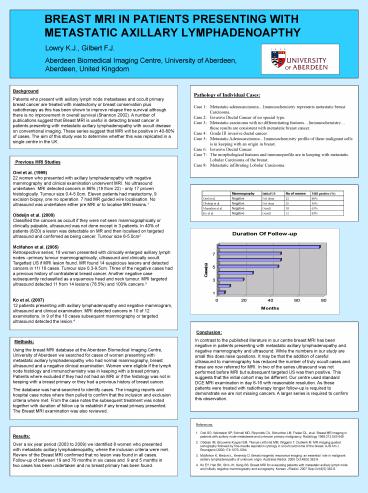
Previous MRI Studies Orel et al. (1999) 22
women who presented with axillary lymphadenopathy
with negative mammography and clinical
examination underwent MRI. No ultrasound
undertaken. MRI detected cancers in 86 (19 from
22) - only 17 proven histologically. Tumour size
0.4-5.0cm. Eleven patients had mastectomy, 9
excision biopsy, one no operation. 7 had MR
guided wire localisation. No ultrasound was
undertaken either pre MRI or to localise MRI
lesions.1 Obdeijn et al. (2000) Classified the
cancers as occult if they were not seen
mammographically or clinically palpable,
ultrasound was not done except in 3 patients. In
40 of patients (8/20) a lesion was detectable on
MR and then localised on targeted ultrasound and
confirmed as being cancer. Tumour size 6-5.0cm2
McMahon et al. (2005) Retrospective series 18
women presented with clinically enlarged axillary
lymph nodes primary tumour mammographically,
ultrasound and clinically occult. Targetted US if
MRI lesion found. MR found 14 suspicious lesions
and detected cancers in 11/ 18 cases. Tumour size
0.3-9.5cm. Three of the negative cases had a
previous history of contralateral breast cancer.
Another negative case subsequently reclassified
as a squamous head and neck tumour. MRI targeted
ultrasound detected 11 from 14 lesions (78.5)
and 100 cancers.3 Ko et al. (2007) 12 patients
presenting with axillary lymphadenopathy and
negative mammogram, ultrasound and clinical
examination. MRI detected cancers in 10 of 12
examinations. In 9 of the 10 cases subsequent
mammography or targeted ultrasound detected the
lesion.4
Conclusion In contrast to the published
literature in our centre breast MRI has been
negative in patients presenting with metastatic
axillary lymphadenopathy and negative mammography
and ultrasound. While the numbers in our study
are small this does raise questions. It may be
that the addition of careful ultrasound to
mammography has reduced the number of truly
occult cases and these are now referred for MRI.
In two of the series ultrasound was not performed
before MRI but subsequent targeted US was then
positive. This suggests that the initial cohort
may be different. Our centre used standard DCE
MRI examination in day 6-16 with reasonable
resolution. As these patients were treated with
radiotherapy longer follow-up is required to
demonstrate we are not missing cancers. A larger
series is required to confirm this observation.
Methods Using the breast MRI database at the
Aberdeen Biomedical Imaging Centre, University of
Aberdeen we searched for cases of women
presenting with metastatic axillary
lymphadenopathy who had normal mammography,
breast ultrasound and a negative clinical
examination. Women were eligible if the lymph
node histology and immunochemistry was in keeping
with a breast primary. Patients where excluded if
they had not had an MRI or if the histology was
not in keeping with a breast primary or they had
a previous history of breast cancer. The database
was hand-searched to identify cases. The imaging
reports and hospital case notes where then pulled
to confirm that the inclusion and exclusion
criteria where met. From the case notes the
subsequent treatment was noted together with
duration of follow up to establish if any breast
primary presented. The Breast MRI examination was
also reviewed.
References
- Orel SG, Weinstein SP, Schnall MD, Reynolds CA,
Schuchter LM, Fraker DL, et al. Breast MR imaging
in patients with axillary node metastases and
unknown primary malignancy. Radiology
1999212543-549 - Obdeijn IM, Brouwers-Kuyper EM, Tilanus-Linthorst
MM, Wiggers T, Oudkerk M. MR imaging-guided
sonography followed by fine-needle aspiration
cytology in occult carcinoma of the breast. AJR
Am J Roentgenol 20001741079-1084. - McMahon K, Medoro L, Kennedy D. Breast magnetic
resonance imaging an essential role in
malignant axillary lymphadenopathy of unknown
origin. Australas Radiol. 2005 Oct49(5)382-9. - Ko EY, Han BK, Shin JH, Kang SS. Breast MRI for
evaluating patients with metastatic axillary
lymph node and initially negative mammography and
sonography. Korean J Radiol. 2007
Sep-Oct8(5)382-9.
Results Over a six year period (2003 to 2009)
we identified 8 women who presented with
metastatic axillary lymphadenopathy, where the
inclusion criteria were met. Review of the Breast
MRI confirmed that no lesion was found in all
cases. Follow-up of between 19 and 76 months in
six cases and 9 and 5 months in two cases has
been undertaken and no breast primary has been
found.
Recommended
CrystalGraphics Presentations


