
Ch 23: Urinary System - PowerPoint PPT Presentation
1 / 27
Title:
Ch 23: Urinary System
Description:
Identify the blood vessels that supply blood to the nephrons ... Uriniferous Tubule p 691. Nephron Collecting Duct (tubule) Renal Corpuscle. PCT. LOH ... – PowerPoint PPT presentation
Number of Views:45
Avg rating:3.0/5.0
Title: Ch 23: Urinary System
1
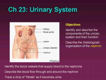
Ch 23 Urinary System
Objectives Identify and describe the components
of the urinary system and their function Describe
the (histological) organization of the nephron
Identify the blood vessels that supply blood to
the nephrons Describe the blood flow through and
around the nephron Trace a drop of filtrate as
it becomes urine
2
Functions of Urinary System (Kidneys)
- Regulate fluid balance (fluid volume) of the body
- Excrete organic waste products and conserve
nutrients, etc - Stabilize pH
- Regulate electrolyte concentrations in the blood
- Endocrine functions
3
Kidney Location
- Lateral to vertebral column high on body wall,
under floating ribs, in retro-peritoneal position
(posterior to the parietal peritoneum) - The right kidney is slightly inferior to the left
kidney in order to accommodate the liver - Surrounded by the renal capsule with a fat pad
- 12 x 6 x 3 cm
- Bean shaped
- Hilus indentation
4
Internal Anatomy
- Cortex outer layer, light reddish brown,
granular appearance (due to many corpuscles) - Medulla darker striped appearance (due to
tubules) Subdivided into distinct renal
pyramids, terminating with a papilla. Separated
by renal columns from the cortex. - Pelvis Expanded proximal ureter
Compare to Fig 23.3
5
(No Transcript)
6
Renal Circulation20-25 of cardiac output!!
7
R
L
8
Nephron functional unit
(gt106/kidney)
- Renal corpuscle
- Glomerulus
- Bowmans (renal) capsule
- Nephron corpuscle
- PCT
- LOH
- DCT
Fig 23.6
9
Renal Corpuscle
10
Uriniferous Tubule p 691
- Nephron Collecting Duct (tubule)
- Renal Corpuscle
- PCT
- LOH
- DCT
- CD
11
This diagram has an important inaccuracy!
12
See Fig 23.4
13
Two Types of Nephrons
Fig 23.9
- Cortical nephrons (85) shorter, mostly in cortex
of kidney, produce "standard" urine - Juxtamedullary nephrons (15), "juxta next to"
the medulla - responsive to ADH, can produce
concentrated urine due to longer Loops of Henle
14
Filtration Passage across Three Barriers
- 1. Capillary endothelium
- Fenestrated
- What gets through?
- 2. Basement membrane
- 3. Glomerular epithelium ( visceral layer of
Bowmans capsule)slit pores between pedicles of
podocytes - Note Capsular Epithelium is simple squamous
epithelium
15
Juxtaglomerular (JG) Apparatus
- Macula densa
- Juxtaglomerular cells (smooth muscle fibers from
afferent arteriole) - Juxtaglomerular Apparatus
- Endocrine system structure (renin and EPO)
16
(No Transcript)
17
Urine collection
- Collecting ducts within each renal papilla
release urine into minor calyx ? major calyx ?
renal pelvis ? ureter
18
Ureters
- From kidney to bladder
- Enter the bladder at an angle
- Trigone
- Retroperitoneal
- Transitional Epithelium
- Nephroliths
This is another inaccuracy!!
19
Urine Transport, Storage, and Elimination
- Trace drop of urine from the afferent arteriole
to the outside world
20
Nephrolithiasis
Occurs when urine becomes too concentrated and
substances crystallize. Symptoms arise when
stones begin to move down ureter causing intense
pain.
Kidney stones may form in the pelvis or calyces
of the kidney or in the ureter.
21
Urinary Bladder
- Retroperitoneal, behind pubis
- Internal folds - rugae - permit expansion (max.
holding capacity 1L) - Trigone - area at base delineated by openings of
ureters and urethra - without muscle - Internal urethral sphincter - involuntary
sphincter - Histology
- 1. transitional epithelium
- 2. detrusor muscle smooth muscle
22
Urinary Bladder
23
Transitional Epithelium
from renal pelvis to neck of urethra.
full bladder
empty bladder
24
Female Urethra
- External urethral sphincters voluntary at
pelvic floor - 3-5 cm from base of bladder to vestibule
- UTIs (esp. E.coli)
25
Male Urethra
- Male 18-20 cm
- 1. prostatic urethra from base of bladder
through prostate gland - 2. membranous urethra between prostate gland
base of penis - 3. penile (spongy) urethra traverses penis to
orifice
26
Male vs. Female
Fig 23.17
27
- Kidneys may sustain 90 loss of nephrons and
still not show apparent symptoms!!! - 2-4 of population only have 1 kidney!
Manneken Pis Fountain Brussels, 1619
28
(No Transcript)
Recommended
CrystalGraphics Presentations


