
Ultrasound - PowerPoint PPT Presentation
1 / 22
Title:
Ultrasound
Description:
US is acoustic vibrations (sound waves) above the frequency audible to the human ... They may experience a deep aching. This is caused by periosteal vibration. ... – PowerPoint PPT presentation
Number of Views:55
Avg rating:3.0/5.0
Title: Ultrasound
1
Ultrasound
2
ULTRASOUND
- Based on principles discovered by Wood and Loomis
in 1927. - US is acoustic vibrations (sound waves) above the
frequency audible to the human ear. - Human ear can detect up to 20,000 cycles per
second. Anything above that is considered
ultrasonic.
3
- Crystal is given electrical impulse from a
transducer causing it to vibrate at a certain
frequency(same as in your watch) - Frequency of vibration is measured in MHz -
usually 1 or 3 MHz - 1 MHz penetrates more deeply,
- 3 MHz more superficial
- the 3 MHz produces more energy which is absorbed
more readily, thus depth is reduced - This vibration causes sound waves which are
directed by US head
4
(No Transcript)
5
Actions of Ultrasound
- Generation of Heat through friction
- heat is deep
- Mechanical actions attributed to high alternating
pressure forces and accelerations
- Vibratory effect which penetrates deeply
6
EQUIPMENT
- This causes a crystal transducer to vibrate and
generate sound waves
- A generator produces electrical oscillations of a
desired frequency
- The sonic energy is transmitted to human tissue
by contact with the ultrasound head
7
Intensity
- Varies between 0 to 3 W/CM2
- Most machines have Watts (W) or W/CM2
- 1-1.5 W/CM2 is typical intensity
8
Types of patients treated
- Muscle injuries
- Tendon injuries
- Ligament injuries
- Other soft tissue injuries
- Inflammatory conditions such as CTS,
Tenosynovitis, DeQuervains, Radial Tunnel Syndrome
9
OTHER CONDITIONS
- Lateral epicondylitis
- Medial epicondylitis
- Muscle spasms
- Do not apply high intensity US (1.5 W/cm2 ) to
acute inflammatory conditions since it tends to
aggravate the condition
10
Continuous Vs. Pulsed US
- Pulsed delivers the ultrasound waves
intermittently - Usually represented in of continuous
- also called duty cycle
- 50, 20
- May be used for tissue healing
- 20, .1 - .2 W/CM2 , 30-60 seconds for each area
the size of transducer head - treatment repeated qd or bid
11
CONTRAINDICATIONS FOR US
- Pregnancy (always ask document)
- Cancer (always ask document)
- Diminished circulation
- Electrical internal or external implanted device
- Spinal cord stimulators
- Acute inflammatory conditions
- Tendon lacerations for 1st 6 weeks
12
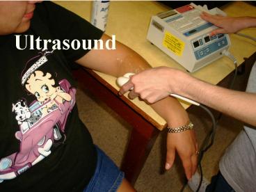
US TECHNIQUE
- Explain to patient how it will feel
- Protect clothing from gel
- Apply US gel to skin liberally
- Spread gel over area to be treated
- Turn intensity knob all the way down
- Set machine to pulsed of continuous
- Set desired treatment time
- Place transducer on skin at right angle to skin.
13
WHAT TO TELL PATIENT ABOUT US
- Explain how it works
- Ask them to tell you immediately if they feel
anything - They may feel warmth. This is normal.
- It should not feel hot. If it does, turn down
the intensity or discontinue the treatment. - They may experience a deep aching. This is
caused by periosteal vibration. Turn down
intensity if this happens.
14
APPLICATION CONTINUED
- Up to 2 W/cm2 in obese pt as long as no C/O heat.
- Use a circular motion.
- Treatment usually lasts 8-15 minutes
- the area that can be effectively treated is 2X
the size of transducer - Move head 1 inch per second
- dont apply if bone blocks pathway
- Switch control to W/cm2
- Begin moving transducer head constantly and
slowly - Slowly turn up intensity
- Intensity usually W/cm2
- Consider the fact that the deeper the tissue, the
poorer the therapeutic dosage due to the
dissipating effect of blood flow
15
Wound Care
- low intensity US promotes healing
- .1-.5 W/cm2
- duty cycle 20
- intensities of .8 W/cm2 or higher have no effect
or retard healing
16
PHONOPHORESIS
- Technique is same as Ultrasound
- Cortisone cream used
- Ultrasonic energy may push cortisone under skin
for anti-inflammatory effect - Or, US may increase cell permeability
- 5-10 cortisone cream used
17
NEVER !!!
- Lift the transducer off the pts skin with the
intensity on - Stop moving the transducer during a treatment
- Do US over bone
- Place transducer directly on the skin
18
UNDERWATER TECHNIQUE
- Transducer must be waterproof
- Useful for irregular surfaces such as a finger,
the hand or the ankle - Place the body part to be treated under water
- Hold transducer 1/2 inch from skin
- Move transducer as you would normally
- Never place directly on skin
- Do not allow patient to touch any sources of
electricity during treatment
19
BE CAREFUL IF...
- Patient has decreased sensation
- Skin is not intact (avoid these areas)
20
Cavitation
- Cavitation - behavior of a micron sized gas
bubble in fluids when energized by US - If intensity too high duration is long enough,
bubbles become progressively larger - They take in air during rarefaction phase than
they loose in compression phase - If bubbles reach critical size, they collapse
under pressure sending shock waves through
tissue. - This is called unstable cavitation
- Air bubbles in water should be avoided
21
- Energy is absorbed in proportion to density
- Periosteum is dense and heats quickly
- bone, ligament and tendon circulation is not as
good as muscle and they retain heat longer
22
NOW LETS PRACTICE
Recommended
CrystalGraphics Presentations


