
Eukaryotic transcription' - PowerPoint PPT Presentation
1 / 12
Title:
Eukaryotic transcription'
Description:
Genomic clones are used to study promoters. and ... Genomic clone. The precise location of a promoter is located by. deletion analysis. Transcription ... – PowerPoint PPT presentation
Number of Views:92
Avg rating:3.0/5.0
Title: Eukaryotic transcription'
1
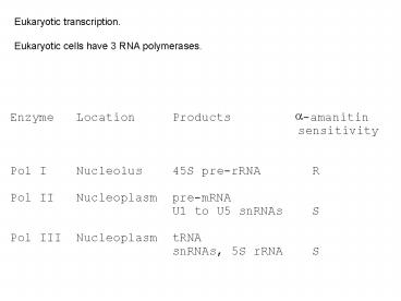
Eukaryotic transcription. Eukaryotic cells have
3 RNA polymerases.
Pol I Nucleolus 45S pre-rRNA R
2
Amanita phalloides , the death cap, synthesises
a-amanitin.
3
Subunit structure of eukaryotic RNA polymerases
Related to bacterial ? subunit. Binds DNA. Has
CTD in Pol II.
200kDa
Related to bacterial ?? subunit. Binds NTPs
100kDa
Related to bacterial ? subunit.
50kDa
3 of the smaller subunits are common to all 3
eukaryotic RNA polymerases
4
All 3 eukaryotic RNA polymerases have 10
subunits.
Some of these are homologous to subunits of
bacterial RNA polymerase ?2 ? ? ? .
The largest RNA pol II subunit has a unique
C-terminal domain (CTD) - multiple (26 to 50)
repeats of the AA sequence YSPTSPS. The CTD has
a role in transcription initiation.
3 subunits appear in all 3 eukaryotic RNA
polymerases.
All 3 eukaryotic RNA polymerases need
additional transcription factor (TF) proteins to
bind promoters and start transcription.
5
Transcription proceeds in the 5 to 3 direction.
RNA polymerases generally dont proof-read.
6
Investigation of transcription in eukaryotes.
Genomic clones are used to study promoters and
transcription initiation.
Genomic clone
cDNA clone
7
Genomic clone
5
3
Exon
Exon
Exon
Promoter should be around here
The ability of upstream DNA to promote
transcription is measured. e. g. by measuring
the amount of RNA transcript produced in an in
vitro transcription system.
8
The precise location of a promoter is located by
deletion analysis.
The transcription start point is identified by
analysing the RNA transcript.
9
Site-directed mutagenesis can be used to
identify important bases in a promoter region.
Up mutations increase promoter activity. Down
mutations decrease promoter activity.
Footprinting is used to identify protein binding
sites in DNA e. g. TF binding sites within
promoters.
10
32P
DNA footprinting
Gel electrophoresis
Autoradiography
11
Gel electrophoresis
Autoradiography
Footprint
12
Control
Bound protein
Real example
Footprint
Increasing concentration of DNA binding protein.
Recommended
CrystalGraphics Presentations


