
The Microscope - PowerPoint PPT Presentation
1 / 24
Title:
The Microscope
Description:
Part of microscope to which the objectives are attached ... A secure part of the microscope to hold on to when the microscope is being carried. ... – PowerPoint PPT presentation
Number of Views:69
Avg rating:3.0/5.0
Title: The Microscope
1
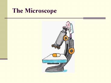
The Microscope
2
The Microscope
- Simple Microscope one lens
- Ex Magnifying lens
- Compound Microscope 2 lenses that compound or
magnify each other. - Dissecting Microscope no special slide
preparation
3
Microscopes
4
Parts of The Compound Microscope
5
Continued
- Eyepiece
- Magnifies material being viewed by 10X
- The part of the microscope you look into
- Sometimes contains a pointer that can be seen as
you look into the eyepiece. - May also be called the ocular.
6
Continued
- Nose piece
- Part of microscope to which the objectives are
attached - Rotates to allow for the changing of objectives
to increase or decrease magnification.
7
Continued
- Arm
- A secure part of the microscope to hold on to
when the microscope is being carried.
8
Continued
- Objectives
- Low (4x)
- Medium (10x)
- High (40x)
9
Continued
- Stage
- Platform on which microscope slide rests
- Mechanical Slide Adjuster
- Used for adjusting the position of the slide for
viewing
10
Continued
- Coarse adjustment knob
- Large movements of the stage
- Fine adjustment knob
- Precise focusing under High power
11
Continued
- Diaphragm
- regulates the amount of light passing through the
slide
12
Continued
- Illuminator
- Light source
- Base
- provides support for microscope
13
Continued
- Body tube
- Connects Ocular to Nosepiece
14
TOTAL MAGNIFICATION
- Power of the eyepiece (10X) multiplied by
objective lenses determines total magnification.
15
Magnification
16
Field of View
- Field of View (FV) is the illuminated circle that
you see when looking through the ocular eyepiece.
- If we know the diameter of the FV then we can
estimate the size of our microorganisms.
17
Field of View
- With our microscopes the diameter of the FV under
low power is 4 mm - FV is measured in micrometers or microns.
- 1 mm 1000 microns
- Therefore, our FV under low power is 4000 microns
18
Using the Field of View to Estimate Microscopic
Measurements
If an organism takes up ½ of the FV under low
power, it must be about 2000 microns in length
19
How does the FV change as Magnification goes Up??
- As magnification goes up, the size of the FV gets
smaller. - If magnification increases 2x, the FV is divided
by 2x, or 2x smaller. - If we switch from low power (4X) to medium power
(10X), the increase in magnification is 2.5 times
(10X/4X). - The FV under medium power will be 1600 microns
(4000/2.5)
20
Calculating the Diameter of the Field of View
- Step 1 Calculate the Increase in Magnification.
New Objective - Old Objective
- Step 2 Divide the old F of V by the increase in
magnification calculated in Step 1 - Old F of V
(Microns) - Increase in Mag
21
To Calculate the Changing FV
- Low 5x Med 10x High 50x
- Low FV 5mm (1000um x 5mm 5000um)
- Step 1 Calculate the increase in Magnification
- Ex 10x/5x 2
- Step 2 Calculate the reduction of the FV
- 5000um/2 2500um
22
Lets try High Power
- Step 1-
- Step 2
50X
23
(No Transcript)
24
Electron Microscopy
- SEM
- TEM