
Novel sdAb Development - PowerPoint PPT Presentation
Title:
Novel sdAb Development
Description:
Creative Biolabs is one of the well-recognized experts who are professional in supporting a broad range of single domain antibody (sdAb) development projects. Along with over a decade of extensive experience in developing novel sdAbs, our scientists are proud to tailor and conduct the best-fit proposal to meet your specific project requirements. – PowerPoint PPT presentation
Number of Views:18
Title: Novel sdAb Development
1
Case Study - SdAb Development for 3 Targets via 1
Camelid Immunization
Single Domain Antibody
Introduction
Single domain antibody (sdAb), is a kind of
antibody fragments
Creative Biolabs has been a long-term expert in
the field of
consisting of a single monomeric variable
antibody domain and lacking the light chain and
CH domain of the heavy chain in conventional Fab
region. In terms of only 12-15 kDa molecular
weight, which is much smaller than either full
length antibody (150-160 kDa) or other antibody
fragments (Fab 50 kDa, scFv 25 kDa), sdAb takes
great advantages of stability and
penetrability, which are essential to the
development of several antibody drugs or
diagnostic tools.
single domain antibody (sdAb) development. Our
scientists have extensive experience in
immunizing camelid animals with the target of
interest to generate novel sdAbs. In terms of our
advanced Hi-Affi phage display platform, we can
use 1 immunized host animal to generate
high-specific sdAbs for multiple antigens. This
is a cost-effective and time-saving option for
specific sdAb development, especially when you
need to investigate different targets with low
homology.
Project Objective Achievement
For this case study, THREE different targets were
provided as antigens and screening targets.
Creative Biolabs is entrusted to immunize only
ONE camelid host animal with these targets and
then develop antigen-specific single domain
antibodies, respectively.
For Target 1, all the 40 clones were observed as
positive through monoclonal phage ELISA and 7
unique VHH sequences have been identified and
confirmed to recognize the target
specifically. For Target 2, all the 40 clones
were observed as positive through monoclonal
phage ELISA and 5 unique VHH sequences have
been identified and confirmed to recognize the
target specifically. For Target 3, 22 of the 40
clones were observed as positive through
monoclonal phage ELISA and 19 unique VHH
sequences have been identified and confirmed
to recognize the target specifically.
With the provided antigens (namely Target 1,
Target 2, and Target 3 or T1, T2, T3 for
short), one camelid was immunized with mixed
antigens. Promising immune response for each
antigen was observed after 4 injections, which
is qualified for library construction. One
uniform immune library was then constructed with
the capacity of over 109. Three rounds of
biopanning were successfully performed against
each of the three targets respectively with
significant good enrichment. 40 clones were
randomly picked from the 3rd round enriched pool
of each target for validation.
Finally, there are 7 unique T1-specific sdAbs, 5
unique T2-specific sdAbs, and 19 unique
T3-specific sdAbs be discovered in this project.
Milestone Overview
Stage 1 Animal Immunization
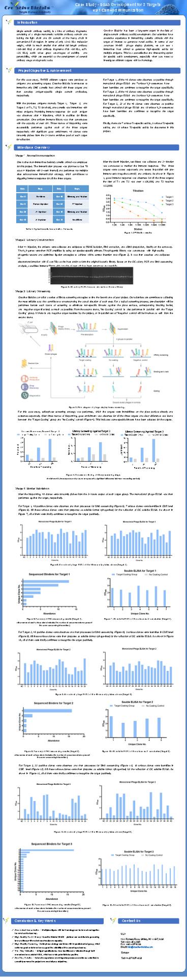
After the fourth injection, test bleed was
collected and 2nd titration was conducted to
monitor the immune response. The three targets
were coated separately and tested in-parallel
with pre- immune sera (negative control) and
antisera. As shown in Figure 1, good immune
response was observed for all the three targets
the titer of T1 and T3 was over 1128,000, and T2
reached 132,000.
One native (non-immunized before) camelid animal
was employed for this project. The immunization
process was planned to last 70 days (4
injections with 3-week interval) and performed
via multiple sites subcutaneous immunization
strategy, which contributes to triggering immune
response for all the three targets.
Date Steps Date Steps
Day 0 Pre-bleed Day 49 Bleeding and Titration
Day 0 Primary Injection Day 63 4th Injection
Day 21 2nd Injection Day 70 Bleeding and Titration
Day 42 3rd Injection Day 72 Final Bleed
Table 1. Typical Camelid Immunization Schedule.
Figure 1. 2nd titration results.
Stage 2 Library Construction After 4th
injection, the antisera were collected and
subjected to PBMC isolation, RNA extraction, and
cDNA preparation, freshly on the same day. The
VHH genes were then PCR amplified by using our
species-specific primers. The phagemid library
was constructed with high-quality phagemid
vectors and optimized ligation strategies to
achieve 100 correct insertion rate (Figure 2).
It was then desalted and subjected
to electrotransformation with E. coli TG1 as the
host strain to form the original bacteria
library. Based on the QC colony PCR and DNA
sequencing analysis, a qualified immune library
with capacity of over 109 has been generated
successfully.
Figure 2. QC colony PCR of random clones from
the end library Stage 3 Library
Screening Creative Biolabs can tailor a series of
library screening strategies to find the best-fit
one of your project. Our scientists are committed
to collecting the most reliable data that
contribute to understanding the actual situation
of each step. For a typical screening process,
pre-absorption will be performed before each
round of screening to eliminate non-specific
binders against the plate surface, corresponding
blocking buffer, and negative target (if exists)
as much as possible. From the second round, No
Coating control is also performed in parallel
with the Target Coating group. If there is
any negative target required by the project, an
in-parallel test of Negative control will be
involved as well from the second round.
Figure 3. Flow diagram of phage display-based
screening. For this case study, solid-phase
screening strategy was performed, which the
targets were immobilized on the plate surface
directly and screened separately. After three
rounds of biopanning, good enrichment was
observed for all the three targets and clear
difference was found between the Target
Coating group and No Coating control (Figure
4). This indicated some specific binders have
been selected for the targets.
Figure 4. Process monitoring of library screening
stage. (Enrichment is increased round by round
and presents significant difference between no
coating control.)
Stage 4 Binder Validation After the biopanning,
40 clones were randomly picked from the 3rd round
output of each target group. The monoclonal phage
ELISA was then performed against the target,
respectively. For Target 1, 40 positive clones
were observed and then processed for DNA
sequencing (Figure 5). 7 unique clones were
identified in CDR level (Figure 6). All these
unique clones were then prepared as soluble
format (phage-free) for the validation of QC
soluble ELISA. As shown in Figure 7, all of them
were finally confirmed to recognize the target
positively.
Figure 5. Monoclonal phage ELISA of the 40
randomly picked clones Target 1.
Figure 6. Summary of DNA sequencing results
Target 1. (Abundance of each unique clone
indicates the number of sequenced clones present
the same sequencing information.)
Figure 7. QC soluble ELISA of the unique sdAb
candidates Target 1.
For Target 2, 40 positive clones were observed
and then processed for DNA sequencing (Figure 8).
5 unique clones were identified in CDR level
(Figure 9). All these unique clones were then
prepared as soluble format (phage-free) for the
validation of QC soluble ELISA. As shown in
Figure 10, all of them were finally confirmed to
recognize the target positively.
Figure 8. Monoclonal phage ELISA of the 40
randomly picked clones Target 2.
Figure 9. Summary of DNA sequencing results
Target 2. (Abundance of each unique clone
indicates the number of sequenced clones present
the same sequencing information.)
Figure 10. QC soluble ELISA of the unique sdAb
candidates Target 2.
For Target 3, 22 positive clones were observed
and then processed for DNA sequencing (Figure
11). 19 unique clones were identified in CDR
level (Figure 12). All these unique clones were
then prepared as soluble format (phage-free) for
the validation of QC soluble ELISA. As shown in
Figure 13, all of them were finally confirmed to
recognize the target positively.
Figure 11. Monoclonal phage ELISA of the 40
randomly picked clones Target 3.
Figure 12. Summary of DNA sequencing results
Target 3. (Abundance of each unique clone
indicates the number of sequenced clones present
the same sequencing information.)
Figure 13. QC soluble ELISA of the unique sdAb
candidates Target 3.
Conclusion Key Words
Contact Us
- One Animal Immunization - Multiple antigens with
low homology can be immunized together for novel
sdAb discovery. - High-Quality SdAb Library - Creative Biolabs
Hi-Affi platform can contribute to generating
immune library with maximized diversity and
capacity. - High Fidelity Screening - Solid-phase strategy
combined with in-parallel control group, which
achieved great enrichment and support the
reliability of the screening outcomes. - Two-Step Validation - Antigen-specific clones
were identified and validated through both
monoclonal and soluble ELISA, which can avoid
potential false positive. - One-Stop Solution - Extensive experience and
integrated procedure enable our scientists to
smoothly advance the project and meet all your
objectives.
USA
45-1 Ramsey Road, Shirley, NY 11967, USA Tel
1-631-381-2994 Fax 1-631-207-8356 Email
info_at_creative-biolabs.com
Europe
Tel 44-207-097-1828
Recommended
CrystalGraphics Presentations


