
AP BIOLOGY Chapter 8 Membrane Structure - PowerPoint PPT Presentation
1 / 31
Title:
AP BIOLOGY Chapter 8 Membrane Structure
Description:
Paramecium have a specialized organelle, the contractile vacuole, that functions ... Contractile Vacuole in a Paramecium. Facilitated Diffusion ... – PowerPoint PPT presentation
Number of Views:589
Avg rating:3.0/5.0
Title: AP BIOLOGY Chapter 8 Membrane Structure
1
AP BIOLOGYChapter 8Membrane Structure
Function
2
Membrane Structure
- The plasma membrane separates the living cell
from its nonliving surroundings. - Thin barrier, 8 nm thick, controls traffic into
and out of the cell. - Selectively permeable, allowing some substances
to cross more easily than others. - Made of lipids, proteins, and carbohydrates
- Made of mostly amphipathic phospholipids (has a
hydrophilic head and hydrophobic tail) - Arranged in the fluid mosaic model with a fluid
phospholipid bilayer embedded with proteins
3
Membrane Models (A History)
- 1895-Charles Overton membranes are made of
lipids because substances that dissolve in lipids
enter cells faster than those that are insoluble - 1917-Irving Langmuir phospholipids dissolved in
benzene would form a film on water when the
benzene evaporated? hydrophilic heads were in
the water - 1925-E. Gorter F. Grendel cell membranes must
be a phospholipid bilayer two molecules thick. - molecules in the bilayer are arranged with the
hydrophobic fatty acid tails are sheltered from
water while the hydrophilic phosphate groups
interact with water.
4
Membrane Models (A History)
- 1935-H. Davson J. Danielli sandwich model in
which the phospholipid bilayer lies between two
layers of globular proteins. - Early images from electron microscopes seemed to
support the Davson-Danielli model and until the
1960s, it was the dominant model. - Investigations revealed two problems
- not all membranes were alike, but differed in
thickness, appearance, and percentage of
proteins. - Second, measurements showed that membrane
proteins are actually not very soluble in water,
but are amphipathic, with hydrophobic and
hydrophilic regions.
5
Membrane Models (A History)
- 1972-S.J. Singer G. Nicolson revised model
membrane proteins are dispersed and individually
inserted into the phospholipid bilayer. - In this fluid mosaic model, the hydrophilic
regions of proteins and phospholipids are in
maximum contact with water and the hydrophobic
regions are in a nonaqueous environment. - A specialized preparation technique,
freeze-fracture, splits a membrane along the
middle of the phospholid bilayer prior to
electron microscopy. - This shows protein particles interspersed with a
smooth matrix, supporting the fluid mosaic
model.
6
Membranes are fluid
- Membrane is held together by hydrophobic
interactions (very weak bonds) - Most of the lipids and some proteins can move
laterally within the membrane - Molecules can also flip-flop in the membrane,
switching layers - Lipids move quickly 2µm/sec. Larger proteins
move much slower. Other proteins are held in
place by the cytoskeleton - Proof When cells are fused, proteins on their
surfaces mix together
7
Membrane Fluidity
- Membranes remain fluid as temperature decreases,
until the phospholipids settle into a solid. - To keep a membrane liquid
- Add more unsaturated lipidskinked tails (where
double bonds are) keep tails further apart - Add cholesterol (animals only)- wedges between
tails - Keeps membranes less fluid by restraining
movement at high temperatures - Keeps membranes liquid by hindering the close
packing of lipids at low temperatures
8
Membranes are Mosaics
- Membranes are a collage of different proteins
embedded in the fluid matrix of the lipid bilayer - Proteins determine the membranes function
- Integral Proteins penetrate the hydrophobic
core. Many are transmembrane which completely
span the membrane - Peripheral Proteins are loosely bound to the
surface of the membrane, often connected to the
integral protein - Some proteins on the cytoplasmic side attach by
cytoskeleton. On the outside, to the ECM
9
Figure 8.6 The detailed structure of an animal
cells plasma membrane, in cross section
10
Membranes are Sided
- Membranes have distinct inside and outside faces
- The lipid layers have different compositions
- Each protein has a specific orientation
- Carbohydrates are usually restricted to the
exterior surface - Orientation of the membrane is determined in the
ER where it is assembled - Molecules starting on the inside of the ER end
up on the outside of the membrane
11
Membrane Protein Functions
12
Carbohydrate Functions
- Cell-cell recognitionthe ability for a cell to
distinguish one type of neighboring cell from
another - Sorting cells into tissues during embryo
development - Rejection of foreign cells from the body (immune
system) - Carbs are usually branched oligosaccharides with
fewer than 15 monomers (oligo few) bonded to
either lipids (glycolipids) or to proteins
(glycoproteins) - The diversity of carbs on the cells surface and
their location let them function as markers that
distinguish one cell from another
13
Traffic Across Membranes
- Most important emergent property of the plasma
membrane is its ability to regulate transport
across cellular boundaries - Membranes are selectively permeable because of
their molecular structure - Permeability of a molecule through a membrane
depends on the interaction of that molecule with
the hydrophobic core of the membrane. - Hydrophobic molecules, like hydrocarbons, CO2,
and O2, can dissolve in the lipid bilayer and
cross easily. - Ions and polar molecules pass through but have
difficulty penetrating the hydrophobic core. - This includes small molecules, like water, and
larger critical molecules, like glucose and other
sugars.
14
Transport Proteins
- Proteins can assist and regulate the transport of
ions and polar molecules. - Specific ions and polar molecules can cross the
lipid bilayer by passing through transport
proteins that span the membrane. - have a hydrophilic channel that certain molecules
or ions can use as a tunnel through the membrane. - Others bind to these molecules and carry their
passengers across the membrane physically. - Each transport protein is specific as to the
substances that it will translocate (move).
15
Diffusion
- Diffusion is the tendency of molecules of any
substance to spread out in the available space. - Driven by the intrinsic kinetic energy (thermal
motion or heat) of molecules. - Molecules will cross the membrane until both
solutions have equal concentrations. - At this dynamic equilibrium as many molecules
pass one way as cross in the other direction. - In the absence of other forces, a substance will
diffuse from where it is more concentrated to
where it is less concentrated, down its
concentration gradient. (potential energy) - Each substance diffuses down its own
concentration gradient, independent of the
concentration gradients of other substances. - The diffusion of a substance across a biological
membrane is passive transport because it requires
no energy from the cell to make it happen.
16
Relative Concentrations
- Differences in the relative concentration of
dissolved materials in two solutions can lead to
the movement of ions from one to the other. - The solution with the higher concentration of
solutes is hypertonic. - The solution with the lower concentration of
solutes is hypotonic. - Solutions with equal solute concentrations are
isotonic. - Imagine that two sugar solutions differing in
concentration are separated by a membrane that
will allow water through, but not sugar. - The hypertonic solution has a lower water
concentration than the hypotonic solution. - More of the water molecules in the hypertonic
solution are bound up in hydration shells around
the sugar molecules, leaving fewer unbound
water molecules. - Unbound water molecules will move from the
hypotonic solution where they are abundant to
the hypertonic solution where they are rarer.
17
Osmosis
- The diffusion of water across a selectively
permeable membrane is a special case of passive
transport called osmosis. - Osmosis continues until the solutions are
isotonic. - The direction of osmosis is determined only by a
difference in total solute concentration. - The kinds of solutes in the solutions do not
matter. - This makes sense because the total solute
concentration is an indicator of the abundance of
bound water molecules (and therefore of free
water molecules). - When two solutions are isotonic, water molecules
move at equal rates from one to the other, with
no net osmosis.
18
Cell survival depends on balancing water uptake
and loss
- Organisms have osmotic problems in either a
hypertonic or hypotonic environment and must have
adaptations for osmoregulation to maintain their
internal environment. - The cells of most land animals are bathed in an
extracellular fluid that is isotonic to the
cells. - Paramecium have a specialized organelle, the
contractile vacuole, that functions as a bilge
pump to force water out of the cell. - The cells of plants, prokaryotes, fungi, and some
protists have walls that contribute to the cells
water balance. - In a hypotonic solution
- Animal cells will swell until the elastic wall
opposes further uptake, and then burst (lyse). - Plant cells become turgid, contributes to the
mechanical support of the plant. - In an isotonic solution
- Animal cells have no net movement of water
- A plant cell is flaccid and the plant may wilt.
- In a hypertonic solution
- Both types of cells lose water, shrivel, its
volume shrinks. - Eventually, the plasma membrane pulls away from
the wall, plasmolysis is usually lethal.
19
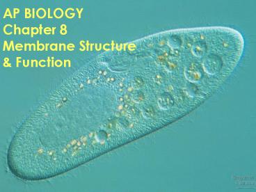
Water Balance in Living Cells
Contractile Vacuole in a Paramecium
20
Facilitated Diffusion
- Many polar molecules and ions that are normally
impeded by the lipid bilayer of the membrane
diffuse passively with the help of transport
proteins that span the membrane. - The passive movement of molecules down its
concentration gradient via a transport protein is
called facilitated diffusion. - Transport proteins
- may have specific binding sites for the solute.
- can become saturated when they are
translocating passengers as fast as they can. - can be inhibited by molecules that resemble the
normal substrate. - they catalyze a physical process, transporting a
molecule across a membrane that would otherwise
be relatively impermeable
21
Type of facilitated diffusion
- Channel proteins serve as corridors allowing a
specific molecule or ion to cross the membrane. - allow fast transport.
- Ex Water channel proteins, aquaporins,
facilitate massive amounts of diffusion. - Some channel proteins, gated channels, open or
close depending on the presence or absence of a
physical or chemical stimulus. - The chemical stimulus is usually different from
the transported molecule. - Ex Neurotransmitters bind to specific gated
channels on the receiving neuron, these channels
open. This allows sodium ions into a nerve cell. - When the neurotransmitters are not present, the
channels are closed. - Some transport proteins do not provide channels
but appear to actually translocate the
solute-binding site and solute across the
membrane as the protein changes shape. - These shape changes could be triggered by the
binding and release of the transported molecule.
22
Active Transport
- Some facilitated transport proteins can move
solutes against their concentration gradient,
from the side where they are less concentrated to
the side where they are more concentrated. - This active transport requires the cell to use
energy (ATP). - Active transport is critical for a cell to
maintain its internal concentrations of small
molecules that would otherwise diffuse across the
membrane. - Active transport is performed by specific
proteins embedded in the membranes. - Adding P from an ATP to the protein induces a
conformational change in the transport protein
that translocates the solute across the membrane.
23
Membrane Potential
- All cells maintain a voltage or membrane
potential across their plasma membranes. - The cytoplasm of a cell is negative in charge
compared to the extracellular fluid - It is due to an unequal distribution of ions on
opposite sides - The membrane potential favors the passive
transport of cations () into the cell and anions
(-) out of the cell. - Two combined forces, collectively called the
electrochemical gradient, drive the diffusion of
ions across a membrane - A chemical force based on an ions concentration
gradient - An electrical force based on the effect of the
membrane potential on the ions movement. - Ions diffuse not simply down their concentration
gradient, but diffuse down their electrochemical
gradient.
24
Sodium Potassium Pump
- The sodium-potassium pump actively maintains the
gradient of sodium (Na) and potassium ions (K)
across the membrane. - Typically, an animal cell has higher
concentrations of K and lower concentrations
of Na inside the cell. - The sodium-potassium pump uses the energy of
one ATP to pump three Na ions out and two K
ions in.
25
Sodium-Potassium Pump, Cont.
- For example Nerve Cell
- Before stimulation there is a higher
concentration of Na outside a resting nerve
cell. - When stimulated, a gated channel opens and Na
diffuses into the cell down the electrochemical
gradient. - Special transport proteins, electrogenic pumps,
generate the voltage gradients across a membrane - The sodium-potassium pump in animals restores the
electrochemical gradient not only by the active
transport of Na and K, but because it pumps two
K ions inside for every three Na ions that it
moves out.
26
Proton Pumps
- In plants, bacteria, and fungi, a proton pump is
the major electrogenic pump, actively
transporting H out of the cell. - Protons pumps in the cristae of mitochondria and
the thylakoids of chloroplasts concentrate H
behind membranes. - These electrogenic pumps store energy that can
be accessed for cellular work.
27
Cotransport
- A single ATP-powered pump that transports one
solute can indirectly drive the active transport
of several other solutes through cotransport via
a different protein. - As the solute that has been actively transported
diffuses back passively through a transport
protein, its movement can be coupled with the
active transport of another substance against its
concentration gradient. - Plants commonly use the gradient of hydrogen
ions that is generated by proton pumps to drive
the active transport of amino acids, sugars,
and other nutrients into the cell. - The high concentration of H on one side of the
membrane, created by the proton pump, leads to
the facilitated diffusion of protons back, but
only if another molecule, like sucrose, travels
with the hydrogen ion.
28
Exocytosis Endocytosis
- Large molecules, such as polysaccharides and
proteins, cross the membrane via vesicles. - Exocytosis
- a transport vesicle budded from the Golgi
apparatus is moved by the cytoskeleton to the
plasma membrane. - When the two membranes come in contact, the
bilayers fuse and spill the contents to the
outside. - Endocytosis
- a cell brings in macromolecules and particulate
matter by forming new vesicles from the plasma
membrane. - A small area of the palsma membrane sinks inward
to form a pocket. - As the pocket into the plasma membrane deepens,
it pinches in, forming a vesicle containing the
material that had been outside the cell. - Endocytosis is a reversal of exocytosis.
29
Types of Endocytosis
- Phagocytosis, cellular eating.
- the cell engulfs a particle by extending
pseudopodia around it and packaging it in a large
vacuole. - the contents are digested when the vacuole fuses
with a lysosome. - Pinocytosis, cellular drinking,
- a cell creates a vesicle around a droplet of
extracellular fluid. - Receptor-mediated endocytosis
- very specific in what substances are being
transported. - This process is triggered when extracellular
substances bind to special receptors, ligands, on
the membrane surface, especially near coated
pits. - triggers the formation of a vesicle.
- enables a cell to acquire bulk quantities of
specific materials that may be in low
concentrations in the environment. - Human cells use this process to absorb
cholesterol. - Cholesterol travels in the blood in low-density
lipoproteins (LDL), complexes of protein and
lipid. - These lipoproteins bind to LDL receptors and
enter the cell by endocytosis. - In familial hypercholesterolemia, an inherited
disease, the LDL receptors are defective, leading
to an accumulation of LDL and cholesterol in the
blood.
30
Types of Endocytosis
31
Figure 8.16 Review passive and active transport
compared
Recommended
CrystalGraphics Presentations


