
Surgical Management of Valve Disease - PowerPoint PPT Presentation
1 / 76
Title:
Surgical Management of Valve Disease
Description:
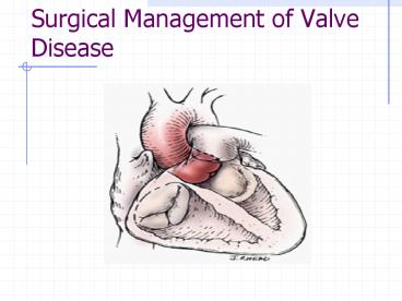
Inflammatory changes affect the aortic valve ... ANGIOGRAM. Mainly indicated in patients over 40yrs to exclude CAD. Identifies Coronary Anomalies ... – PowerPoint PPT presentation
Number of Views:727
Avg rating:3.0/5.0
Title: Surgical Management of Valve Disease
1
Surgical Management of Valve Disease
2
(No Transcript)
3
(No Transcript)
4
Aortic Stenosis
- Obstruction to the blood flow leaving the left
ventricle - Classified into
- Rheumatic--- Pancarditis
- Rheumatic Fever
- Non-Rheumatic Congenital
- Degenerative
5
Physiology
- RHEUMATIC AORTIC STENOSIS
- Least common cause usually associated with mitral
stenosis - Inflammatory changes affect the aortic valve
- Causes scarring and degeneration in the valve
leaflets
6
Physiology
- Resulting in a stenotic valve or destruction of
the soft tissue in the aortic annulus resulting
in dilatation and aortic regurgitation - Decreased incidence in the developed world
7
Degenerative Aortic Stenosis
- Occurs in the normal Aortic Valve
- The Calcific deposit develops as part of the
atheromatous process of the leaflet and causes
the leaflet to remain closed - Developes in the 6th 7th and 8th decades in life
8
Degenerative Aortic Stenosis
- Clinical Risk Factors
- Age
- Male
- Diabetes
- Hypertension
- Renal Failure
- Hyperlipidaemia
- Hypercalcaemia
9
Congenital Bicuspid Valve
- Bicuspid Aortic Valve, the valve has 2 instead of
3 cusps or leaflets - - Is progessive and produces significant
stenosis by the 5th and 6th decades in life - Causes fusion of the commissural leaflet
- Usually the valve orriface is fixed
10
11
Symptoms
- CLASSICAL TRIADE
- Angina
- Syncope
- Congestive Heart Failure
12
ANGINA
- Is the initial symptom in 50-70 of patients
with severe aortic Stenosis - Life Expectancy lt 5 years
- Occurs on exertion
- Angina at rest would be associated with coronary
disease
13
SYNCOPE
- Is the presenting symptom in 15-30 off
presenting Patients - Occurs during exercise as a consequence of
- Reduction of the systematic vascular resistance
without the ability to increase the cardiac
output across the stenotic valve
14
CONGESTIVE HEART FAILURE
- Symptoms include.
- Pulmonary Hypertension.
- Dyspnoea.
- Orthopnoea.
- Paroxismal Noctural Dyspnoea.
- Pulmonary Oedema.
- End Stage Right Ventricular Failure.
15
Cause Of Diastolic Dysfunction.
- Increase LV wall thickness reduces ventricular
compliance - The Atrium needs higher pressure to fill the
ventricle - Development of symptoms of pulmonary congestion
16
CXR
- Normal Heart size with inceased convexity of the
left ventricular silouette due to ventricular
hpertrophy - Calcification of the Aortic Valve
- Cardiomegaly at end stage
17
ECG
- Sign of Left ventricular hypertrophy
- Sign of left atrial enlargement
- Bundle Branch Block
- Complete Heart Block
- Atrial Fibrillation
18
ECHOCARDIOGRAPHY
- Motility of the Leaflet
- Gradient across the valve
- Measure of the valve ares
- EF
- Regional Ventricular Motion
- Ventricular Hypertrophy
- Left Atrial Enlargement
19
ANGIOGRAM
- Mainly indicated in patients over 40yrs to
exclude CAD - Identifies Coronary Anomalies
- Aortogram to identify post stenotic dilatition
- Calcium deposits in the Aortic wall
20
PATHOPHYSIOLOGY
- The natural progresion of Aortic Stenosis is from
- Mild
- Moderate
- Severe
- Development of symptoms indicate
- Moderate Aortic Stenosis
- Critical Aortic Stenosis
21
Moderate Aortic Stenosis
- Valve area of 0.7-1.2cm2
- LV hypertrophy
- Compromised LV contractility causing reduced EF
22
CRITICAL AORTIC STENOSIS
- Valve Area lt0.7cm2
- Increase LVEDP
- Decreased EF
- Decreased Stroke Volume
23
Atrial Fibrillation
- SYMPTOMS MAY APPEAR as ATRIAL CONTRACTION
SUPPLIES UP TO 40 OF THE VENTRICULAR FILLING
DURING DIASTOLE IN AORTIC STENOSIS (20 IN A
NORMAL HEART) - AF MAY CAUSE RAPID DETERIORATION
24
Natural History
- Long Asymptomatic period
- When symptoms develop duration of life is
markedly reduced - After angina occurs survival approx 4yrs After
Syncope approx 2yrs - Congestive Heart Failure Less than 1 yr
25
Natural History
- Valve ares not the only prognostic factor
- Absence of symptoms does not guarantee absence of
L V dysfunction - Sudden Death gt10 in symptomatic Patients
26
Bacterial Endocarditis
- Staphylococcus Aureus most common organism
- Infection most commonly causes valvular
incompetence (regurgitation) from destruction of
the valve and supporting structures - Virulent cases can be seen in IV drug abusers who
use unsterile needles
27
(No Transcript)
28
Pre-op Care
- Diuretics for Heart Failure
- Nitrates and Beta Blockers for Angina
- No proven preventative treatment ? Use of Lipid
Lowering Therapy may slow the progression on
Calcific Aortic Stenosis
29
Indications and Timing of Surgery
- Surgery indicated for all symptomatic patients
- Early detection with regular follow up advised
30
Aortic Regurgitation
- Leakage of Blood from the Aorta back into the
Left Ventricle - Valve fails to close due to damage in the valve
leaflets or changes in the valve annulus - Acute or Chronic
31
Aortic Regurgitation
- Acute
- Infective endocarditis
- Prosthetic Valve Dysfunction
- Chronic
- Dilated Aortic Root
- Rheumatic Fever
- Bicuspid valve
- Hypertension
- Degenerative or inflammatory process (Marfans
syndrome)
32
Aortic Regurgitation
- Large volume of blood is regurgitated into the
Left Ventricle in each diastole - Left Ventricular output may be more than doubled
causing - Left Ventricular Hypertrophy
33
Aortic Regurgitation
- Symptoms
- Dysponea especially on exertion or lying flat
- Fatigue
- Dizziness and awareness of vigorous heart action
- Angina infrequent
34
Aortic Regurgitation
- ECG Evidence of LV Hypertrophy
- CXR LV enlargement ,Dilation of the Ascending
Aorta
35
Aortic Regurgitation
- Clinical Course
- Mild Regurg compatible with normal lifestyle
- Symptom less
- Risk of endocarditis
- Treatment with diuretics and vasodilators for
heart failure
36
Aortic Regurgitation
- Moderate Severe
- Dysponea on exertion or when lying flat
- Fatigue
- May be associated with Aortic Stenosis
37
Aortic Regurgitation
- Chronic Aortic Regurg can be better tolerated
than Aortic Stenosis if compensatory mechanisms
occur - Condition unlikely to improve in the long term
- Surgery should be performed before LV dysfunction
occurs - Acute Aortic Regurg requires emergency surgery
38
(No Transcript)
39
Mitral Stenosis
- The narrowing of the valve orifice which reduces
blood flow leaving the left atrium and entering
the left ventricle. - Left Atrial pressure rises, enlarging the left
atrium causing pulmonary congestion. - Left ventricular filling becomes dependant on
left atrial contraction. - Normal MV area is 4.0-5.0cm2.
40
(No Transcript)
41
Mitral Stenosis
- When valve narrows to lt 2.5cm2 patients become
symptomatic. - Causes.
- Degenerative disease.
- Rheumatic Heart Disease.
42
Degenerative Disease
- Associated with aging
- Diabetes, hypertension and aortic stenosis
- Occurs most often in patients over 70
- Affects women more (41)
- Can appear in 10 of patients over 50
43
Effects
- Calcification occurs where the posterior leaflet
meets the left ventricle and impairs leaflet
mobility - As calcification progresses the annulus becomes
stenotic and blood has a harder time moving
forward through the narrowed valve - Thrombi can form on these areas predisposing
patients to stroke and bacterial endocarditis
44
Rheumatic Heart Disease
- Attributed to Rheumatic Fever which causes
inflammation in various body tissues including
the heart, where the most serious damage occurs - The predominant cause of Mitral Stenosis
45
Causes
- Strep infection, occurs most commonly in children
5-15yrs, less common now in industrialized
nations - Disease follows a stable course initially but in
later years there is a more progressive
acceleration of the disease
46
Effects
- Translucent vegetations appear along the free
margins of the leaflets , causing then to become
inflamed and fuse together - Creating a valve orifice that is stenotic
- Left atrium has difficulty emptying through a
narrow orifice
47
Mitral Stenosis
- Complications
- Atrial Fibrillation an important complication
because it contributes to the development of
Cardiac Failure and is responsible for atrial
stasis risk of thrombosis and embolism - Pulmonary Embolism
- Systemic Embolism cerebral, mesenteric or renal
- Respiratory congestion makes the patient liable
to attacks of acute bronchitis and the
development of chronic bronchitis - Infective endocarditis rare in pure mitral
stenosis, more associated with mixed mitral
stenosis and mitral regurg
48
Mitral Stenosis
- Symptoms
- Symptom free for many years
- Eventually develop features of left sided failure
progressing o right sided failure - Various factors such as Pregnancy or the onset of
Atrial Fibrillation may suddenly precipitate the
patient from one stage to another - Most significant symptom Shortness of Breath
- Strenuous exercise initially progressing to
- Orthopnea
- Paroxysmal Dyspnoea
- Acute Pulmonary Oedema
49
Mitral Stenosis
- ECG A fib common, Evidence of R ventricular
hypertrophy develops in late stages - CXR Classical Feature selective enlargement
of the left atrium - Calcification of the mitral Valve
- Upper pulmonary veins are usually prominent
50
(No Transcript)
51
Mitral Stenosis
- Indications for surgery
- Surgery or Balloon valvuloplasty are eventually
required in most cases - Post relief of the stenosis treatment is still
required for control of arrhythmias and the
prevention of emboli - Balloon Valvuloplasty is useful if the leaflets
are pliant and mobile - Relieve symptoms for 5-10yrs but restenosis
usually occurs
52
Mitral Regurgitation
- Occurs when a valve does not close properly,
causing blood to leak back from the left
ventricle to the left atrium - Increases the workload of the left side to clear
the regurgitated blood - May occur in conjunction with mitral stenosis
53
Causes
- Mitral Valve Prolapsed congenital or acquired
- Congenital disease, rheumatic fever
- Connective tissue disorders Marfans
- Structural factors- disproportion between the
chordae tendane and papillary muscle - Disproportion between mitral leaflet and valve
orifice area often due to cardiomyopathy - More common in young to middle aged women
54
55
Endocarditis
- Infection of the valve by direct invasion of
bacteria - Untreated always fatal
- At risk are patients with congenital valve
defects - Rheumatic heart disease
- 10-15 of nosocomal endocarditis with cardiac
surgery
56
Ischemia
- Complication of Coronary Heart Disease and Acute
MI - Poor perfusion to the papillary muscle
- Necrosis and fibrosis formation causing papillary
muscle dysfunction
57
Effects
- With systole the blood shunts back through the
mitral orifice into the left atrium - Left Atrial pressure rises
- During diastole additional blood returns to the
LV increasing volume
58
Effects
- Both the LA and LV hypertrophy over time
- With LA compensation pulmonary congestion and
dyspnea occur - With LV compensation pulmonary venous pressures
elevate and pulmonary hypertension occur - Ventricular function is compromised and C.O
decreases, consequently L atrial enlargement
causes Afib which further decreases C.O
59
Mitral Rergurgitation
- Acute
- Myocardial ischaemia and Mi
- Endocarditis
- Idiopathic chordae rupture
- Chronic
- Progressive
- Lv dysfunction in spite of normal EF
- patients may remain asymptomatic well after LV
decompensation
60
Mitral Regurgitation
- Indications for surgery
- Acute MR with CCF or Cardiogenic shock
- Acute endocarditis
- CHF due to valvular dysfunction
- Unstable valve prosthesis
- Uncontrolled infection
- Persistent bacterimea
- Fungal infections
- Vegetations in situ
61
Mitral Regurgitation
- Mitral Valve Reconstruction in possible in a
percentage of cases with degenerative disease - Mitral Valve replacement indicated when repair
not possible
62
(No Transcript)
63
64
Types and Choices Prosthetic Valves
- Mechanical
- Better Durability
- Require Long-term Anticoagulation Therapy
- 3 main types
- Ball valves (Starr-Edwards)
- Tilting disc valves (Shiley)
- Bi-leaflet valves (St Jude)
65
Tissue Valves
- Autologous valves made from the patients own
pericardium - Autograft valves diseased aortic valve
removed and replaced with the pulmonary
valve which is then replaced with a
homograft
66
(No Transcript)
67
Tissue Valves
- Homograft Human cadavers
- Porcine Specially treated porcine tissue
- Shorter Lifespan may require replacing within
10yrs - Useful for younger childbearing women
68
(No Transcript)
69
Complications of Prosthetic Valves
- Thromboembolism anticoagulant therapy
- Haemorrhagic Complications
- Primary Valve Failure
- Endocarditis
70
(No Transcript)
71
Pre Operative Considerations
- Nutritional Support
- Aggressive Diuresis
- Measures to prevent Resp Failure
- Atrial Fibrillation
72
Pre Operative Considerations
- Dental Checks
- Cardiac catheterisation
- Selection of valve type
- Counseling
- Asymoathomatic patients question the need for
such dramatic surgery - Valve choice
- Lifestyle changes relating to warfarin therapy
- Education re endocarditis
73
Post-operative Considerations
- Routine complications post cardiac Surgery
- Fluid and haemodynamic monitoring and management
- Education re anticoagulation therapy
- Arrhythmias, complete heart block, need for
permanent pacemaker - Dyspnoea reduction may take time to improve
- Prophylactic antibiotic therapy prior to dental
work and other invasive procedures
74
Antibiotics Prophylaxis
- Dental/oral/resp/oes procedures
- Standard regimen Ampicil 2grm po 1hr
- Unable to take po Ampicil 2grm iv/im 30min
- Pen allergic Clindamycin 600mg or Cefazolin
igrm iv 30mins
75
(No Transcript)
76
(No Transcript)
Recommended
CrystalGraphics Presentations


