
Angular Movement - PowerPoint PPT Presentation
1 / 44
Title: Angular Movement
1
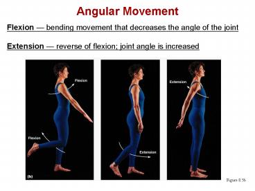
Angular Movement
Flexion bending movement that decreases the
angle of the joint Extension reverse of
flexion joint angle is increased
Figure 8.5b
2
Angular Movement
Dorsiflexion and plantar flexion up and down
movement of the foot Adduction movement toward
the midline Circumduction movement describes a
cone in space
3
Rotation
- The turning of a bone around its own long axis
- Examples
- Between first two vertebrae
- Hip and shoulder joints
Figure 8.5g
4
Special Movements
- Supination and pronation
- Inversion and eversion
- Opposition
5
Muscles Crossing the Shoulder
- Nine muscles cross the shoulder joint and insert
into the humerus - Prime movers include
- Pectoralis major arm flexion
- Latissimus dorsi and posterior fibers of the
deltoid arm extension - Middle fibers of the deltoid arm abduction
6
Muscles Crossing the Shoulder
Posterior
Anterior
Figure 10.14
7
Muscles Crossing the Shoulder
- Rotator cuff muscles supraspinatus,
infraspinatus, teres minor, and subscapularis - Function mainly to reinforce the capsule of the
shoulder - Secondarily act as synergists and fixators
- The coracobrachialis and teres major
- Act as synergists
- Do not contribute to reinforcement of the
shoulder joint
8
Muscles Crossing the Shoulder
Figure 10.14
9
Muscles Crossing the Shoulder
Figure 10.14c
10
Muscles Crossing the Elbow
- Forearm extension
- The triceps brachii is the prime mover of forearm
extension - The anconeus is a weak synergist
- Forearm flexion
- Brachialis and biceps brachii are the chief
forearm flexors - The brachioradialis acts as a synergist and helps
stabilize the elbow
11
Muscles of the Forearm
- The two functional forearm muscle groups are
- those that cause wrist movement, and
- those that move the fingers and the thumb
- These muscles insert via strong ligaments called
flexor and extensor retinacula - Most anterior muscles are flexors, and posterior
muscles are extensors - The pronator teres and pronator quadratus are not
flexors, but pronate the forearm - The supinator muscle is a synergist with the
biceps brachii in supinating the forearm
12
Muscles of the Forearm Anterior Compartment
- These muscles are primarily flexors of the wrist
and fingers
Figure 10.15a
13
Muscles of the Forearm Anterior Compartment
Figure 10.15b, c
14
Muscles of the Forearm Posterior Compartment
- These muscles are primarily extensors of the
wrist and fingers
15
Muscle Action of the Arm Summary
- The posterior extensor and anterior flexor
muscles are shown
16
Muscle Action of the Forearm Summary
- Posterior extensors of the wrist and fingers, and
anterior flexor muscles are shown
17
Intrinsic Muscles of the Hand
- These small muscles
- Lie in the palm of the hand (none on the dorsal
side) - Move the metacarpals and fingers
- Control precise movements (e.g., threading a
needle) - Move the thumb toward the little finger
18
Intrinsic Muscles of the Hand
Figure 10.18a
19
Intrinsic Muscles of the Hand
Figure 10.18b
20
Finger and Thumb Movements
- Flexion
- Thumb bends medially along the palm
- Fingers bend anteriorly
- Extension
- Thumb points laterally
- Fingers move posteriorly
21
Intrinsic Muscles of the Hand Groups
- There are three groups of intrinsic hand muscles
- The thenar eminence (ball of the thumb) and
hypothenar eminence (ball of the little finger)
each have a flexor, an abductor, and an opponens
muscle - The midpalm muscles, the lumbricals and
interossei, extend the fingers - The interossei also abduct and adduct the fingers
22
Intrinsic Muscles of the Hand Groups
Figure 10.18c, d
23
Muscles Crossing Hip and Knee Joints
- Most anterior compartment muscles of the hip and
thigh flex the femur at the hip and extend the
leg at the knee - Posterior compartment muscles of the hip and
thigh extend the thigh and flex the leg - The medial compartment muscles all adduct the
thigh - These three groups are enclosed by the fascia lata
24
Movements of the Thigh at the Hip Flexion and
Extension
- The ball-and-socket hip joint permits flexion,
extension, abduction, adduction, circumduction,
and rotation - The most important thigh flexors are the
iliopsoas (prime mover), tensor fasciae latae,
and rectus femoris - The medially located adductor muscles and
sartorius assist in thigh flexion - Thigh extension is primarily effected by the
hamstring muscles (biceps femoris,
semitendinosus, and semimembranosus) - Forceful extension is aided by the gluteus
maximus
25
Movements of the Thigh at the Hip Flexion and
Extension
Figure 10.19a
26
Movements of the Thigh at the Hip Other
Movements
- Abduction and rotation are effected by the
gluteus medius and gluteus minimus, and are
antagonized by the lateral rotators - Thigh adduction is the role of five adductor
muscles (adductor magnus, adductor longus, and
adductor brevis the pectineus, and the gracilis)
27
Movements of the Thigh at the Hip Other
Movements
Figure 10.20b
28
Movements of the Knee Joint
- The sole extensor of the knee is the quadriceps
femoris - The hamstring muscles flex the knee, and are
antagonists to the quadriceps femoris
Figure 10.19a
29
Fascia of the Leg
- A deep fascia of the leg is continuous with the
fascia lata - This fascia segregates the leg into three
compartments anterior, lateral, and posterior - Distally, the fascia thickens and forms the
flexor, extensor, and fibular retinaculae
Figure 10.22a
30
Muscles of the Leg Movements
- Various leg muscles produce the following
movements at the - Ankle dorsiflexion and plantar flexion
- Intertarsal joints inversion and eversion of
the foot - Toes flexion and extension
31
Muscles of the Anterior Compartment
- These muscles are the primary toe extensors and
ankle dorsiflexors - They include the tibialis anterior, extensor
digitorum longus, extensor hallucis longus, and
fibularis tertius
Figure 10.21a
32
Muscles of the Anterior Compartment
Figure 10.21b-d
33
Muscles of the Lateral Compartment
- These muscles plantar flex and evert the foot
- They include the fibularis longus and fibularis
brevis muscles
Figure 10.22a
34
Muscles of the Lateral Compartment
Figure 10.22b, c
35
Muscles of the Posterior Compartment
- These muscles primarily flex the foot and the
toes - They include the gastrocnemius, soleus, tibialis
posterior, flexor digitorum longus, and flexor
hallucis longus
Figure 10.23a
36
Muscles of the Posterior Compartment
Figure 10.23b, c
37
Muscles of the Posterior Compartment
Figure 10.23d-f
38
Muscle Actions of the Thigh Summary
- Thigh muscles
- Flex and extend the thigh (posterior compartment)
- Extend the leg (anterior compartment)
- Adduct the thigh (medial compartment)
39
Muscle Actions of the Thigh Summary
Figure 10.24a
40
Muscle Actions of the Leg Summary
- Leg muscles
- Plantar flex and evert the foot (lateral
compartment) - Plantar flex the foot and flex the toes
(posterior compartment) - Dorsiflex the foot and extend the toes (anterior
compartment)
41
Muscle Actions of the Leg Summary
Figure 10.24b
42
Intrinsic Muscles of the Foot
- These muscles help flex, extend, abduct, and
adduct the toes - In addition, along with some leg tendons, they
support the arch of the foot - There is a single dorsal foot muscle, the
extensor digitorum brevis, which extends the toes - The plantar muscles occur in four layers
43
Plantar Muscles First Second Layers
- Superficial muscles of the plantar aspect of the
foot - These muscles are similar to the corresponding
muscles of the hand
Figure 10.25a
44
Plantar Muscles Third Fourth Layers
Figure 10.25d