
The cranial nerves are 12 pairs of nerves that can be seen on the ventral bottom surface of the brai - PowerPoint PPT Presentation
1 / 43
Title:
The cranial nerves are 12 pairs of nerves that can be seen on the ventral bottom surface of the brai
Description:
Facial VII. Auditory (vestibulocochlear) VIII. Glossopharyngeal IX. Vagus X. Spinal Accessory XI ... CN VII (Facial), CN IX (Glossopharyngeal) and CN X )Vagus ... – PowerPoint PPT presentation
Number of Views:887
Avg rating:3.0/5.0
Title: The cranial nerves are 12 pairs of nerves that can be seen on the ventral bottom surface of the brai
1
cranial nerve
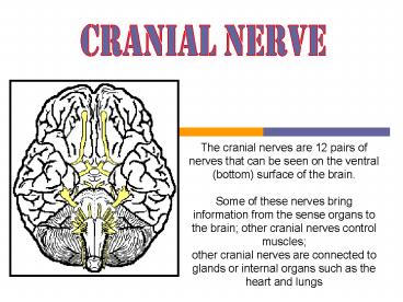
The cranial nerves are 12 pairs of nerves that
can be seen on the ventral (bottom) surface of
the brain. Some of these nerves bring
information from the sense organs to the brain
other cranial nerves control muscles other
cranial nerves are connected to glands or
internal organs such as the heart and lungs
2
To help memorize each, a mnemonic is often used
by students such as . . ."On Old Olympic
Towering Tops A Finn And German Viewed Some Hops"
Olfactory I
Optic II
Oculomotor III
Trochlear IV
Trigeminal V
Abducens VI
Auditory (vestibulocochlear) VIII
Facial VII
Glossopharyngeal IX
Vagus X
Spinal Accessory XI
Hypoglossal XII
3
To help memorize each, a mnemonic is often used
by students such as . . ."On Old Olympic
Towering Tops A Finn And German Viewed Some Hops"
Olfactory I
Optic II
Oculomotor III
Trochlear IV
Trigeminal V
Abducens VI
Auditory (vestibulocochlear) VIII
Facial VII
Glossopharyngeal IX
Vagus X
Spinal Accessory XI
Hypoglossal XII
4
To help memorize each, a mnemonic is often used
by students such as . . ."On Old Olympic
Towering Tops A Finn And German Viewed Some Hops"
Olfactory I
Optic II
Oculomotor III
Trochlear IV
Trigeminal V
Abducens VI
Auditory (vestibulocochlear) VIII
Facial VII
Glossopharyngeal IX
Vagus X
Spinal Accessory XI
Hypoglossal XII
5
To help memorize each, a mnemonic is often used
by students such as . . ."On Old Olympic
Towering Tops A Finn And German Viewed Some Hops"
Olfactory I
Optic II
Oculomotor III
Trochlear IV
Trigeminal V
Abducens VI
Auditory (vestibulocochlear) VIII
Facial VII
Glossopharyngeal IX
Vagus X
Spinal Accessory XI
Hypoglossal XII
6
To help memorize each, a mnemonic is often used
by students such as . . ."On Old Olympic
Towering Tops A Finn And German Viewed Some Hops"
Olfactory I
Optic II
Oculomotor III
Trochlear IV
Trigeminal V
Abducens VI
Auditory (vestibulocochlear) VIII
Facial VII
Glossopharyngeal IX
Vagus X
Spinal Accessory XI
Hypoglossal XII
7
To help memorize each, a mnemonic is often used
by students such as . . ."On Old Olympic
Towering Tops A Finn And German Viewed Some Hops"
Olfactory I
Optic II
Oculomotor III
Trochlear IV
Trigeminal V
Abducens VI
Auditory (vestibulocochlear) VIII
Facial VII
Glossopharyngeal IX
Vagus X
Spinal Accessory XI
Hypoglossal XII
8
To help memorize each, a mnemonic is often used
by students such as . . ."On Old Olympic
Towering Tops A Finn And German Viewed Some Hops"
Olfactory I
Optic II
Oculomotor III
Trochlear IV
Trigeminal V
Abducens VI
Auditory (vestibulocochlear) VIII
Facial VII
Glossopharyngeal IX
Vagus X
Spinal Accessory XI
Hypoglossal XII
9
To help memorize each, a mnemonic is often used
by students such as . . ."On Old Olympic
Towering Tops A Finn And German Viewed Some Hops"
Olfactory I
Optic II
Oculomotor III
Trochlear IV
Trigeminal V
Abducens VI
Auditory (vestibulocochlear) VIII
Facial VII
Glossopharyngeal IX
Vagus X
Spinal Accessory XI
Hypoglossal XII
10
To help memorize each, a mnemonic is often used
by students such as . . ."On Old Olympic
Towering Tops A Finn And German Viewed Some Hops"
Olfactory I
Optic II
Oculomotor III
Trochlear IV
Trigeminal V
Abducens VI
Auditory (vestibulocochlear) VIII
Facial VII
Glossopharyngeal IX
Vagus X
Spinal Accessory XI
Hypoglossal XII
11
To help memorize each, a mnemonic is often used
by students such as . . ."On Old Olympic
Towering Tops A Finn And German Viewed Some Hops"
Olfactory I
Optic II
Oculomotor III
Trochlear IV
Trigeminal V
Abducens VI
Auditory (vestibulocochlear) VIII
Facial VII
Glossopharyngeal IX
Vagus X
Spinal Accessory XI
Hypoglossal XII
12
To help memorize each, a mnemonic is often used
by students such as . . ."On Old Olympic
Towering Tops A Finn And German Viewed Some Hops"
Olfactory I
Optic II
Oculomotor III
Trochlear IV
Trigeminal V
Abducens VI
Auditory (vestibulocochlear) VIII
Facial VII
Glossopharyngeal IX
Vagus X
Spinal Accessory XI
Hypoglossal XII
13
To help memorize each, a mnemonic is often used
by students such as . . ."On Old Olympic
Towering Tops A Finn And German Viewed Some Hops"
Olfactory I
Optic II
Oculomotor III
Trochlear IV
Trigeminal V
Abducens VI
Auditory (vestibulocochlear) VIII
Facial VII
Glossopharyngeal IX
Vagus X
Spinal Accessory XI
Hypoglossal XII
14
To help memorize each, a mnemonic is often used
by students such as . . ."On Old Olympic
Towering Tops A Finn And German Viewed Some Hops"
Olfactory I
Optic II
Oculomotor III
Trochlear IV
Trigeminal V
Abducens VI
Auditory (vestibulocochlear) VIII
Facial VII
Glossopharyngeal IX
Vagus X
Spinal Accessory XI
Hypoglossal XII
15
QUIZ
TEST UR CRANIAL NERVE
16
X. Vagus Nerve
"Vagus" is from the Latin meaning wandering The
vagus nerve consists of fivecomponents with
distinct functions
17
X. Vagus Nerve
Overview of Branchial Motor Component
Origin
from the nucleus ambiguus in the reticular
formation of the medulla.
18
X. Vagus Nerve
Overview of Branchial Motor Component
Pathway
Fibers leaving the nucleus ambiguus travel
anteriorly and laterally to exit the medulla
posterior to the oliveas The branchial motor
component travels with the fibers of (CN XI)
into the jugular foramen of the skull and give
rise to two ganglia (the superior and inferior
vagal ganglia) within the jugular foramen. The
branchial motor fibers join with the rest of the
vagus nerve just below the inferior vagal
ganglion. Upon exiting the skull the vagus nerve
travels between the internal jugular vein and
internal carotid artery within the carotid sheath.
19
X. Vagus Nerve
Overview of Branchial Motor Component
- The branchial motor fibers leave the vagus nerve
as three major branches - Pharyngeal branch
- Superior laryngeal nerve
- Recurrent laryngeal nerve
20
X. Vagus Nerve
Overview of Branchial Motor Component
Action
- Branchial motor component of CN X provides
voluntary control of the - Striated muscle of the pharynx.
- Striated muscle of the larynx, except for
- 1) the stylopharyngeus muscle ..
- 2) the tensor veli palatini muscle ..
- Palatoglossus muscle of the tongue.
21
X. Vagus Nerve
Overview of visceral Motor Component
Origin
from the dorsal motor nucleus of the vagus .
22
X. Vagus Nerve
Overview of visceral Motor Component
Pathway
Upon emerging from the lateral aspect of the
medulla, to exit the skull via the jugular
foramen, the vagus nerve travels between the
internal jugular vein and internal carotid
artery within the carotid sheath Within the
thorax the left and right vagus nerves break up
into many branches to form plexuses around the
esophagus and major blood vessels to the heart
and lungs. From the esophageal plexus, the left
and right gastric nerves emerge and provide
preganglionic parasympathetic innervation to the
stomach, intestines and visceral organs generally
follow the arterial blood supply to that organ.
23
X. Vagus Nerve
Overview of visceral Motor Component
Action
parasympathetic stimulation for the Cardiac
- Slows heart rate Lungs - increased
bronchiolar secretions and bronchoconstriction
GI tract- increased secretions and motility
24
X. Vagus Nerve
Overview of visceral sensory Component
Origin
from solitary nucleus, spinal trigeminal nucleus
inferior ganglion .
25
X. Vagus Nerve
Overview of visceral sensory Component
Peripheral Course
Sensory fibers from the plexuses surrounding the
abdominal viscera and join the gastric nerves
which ascend through the esophageal hiatus of the
diaphragm and merge with the esophageal plexus.
In the thorax, visceral sensory fibers from the
heart and lungs also join the ascending fibers in
the esophageal plexus which converge to form the
left and right vagus nerves which ascend within
the carotid sheath between the internal jugular
vein and internal carotid artery.
Visceral sensory fibers from the larynx and
pharynx join the ascending vagal fibers via the
internal laryngeal and recurrent laryngeal
nerves. these afferent neurons reside in the
inferior vagal ganglion in the jugular foramen.
26
X. Vagus Nerve
Overview of visceral sensory Component
Central Course
The central processes of the visceral sensory
neurons pass from the inferior vagal ganglion
through the jugular foramen and enter the
medulla.
These fibers descend in the tractus solitarius to
synapse in the caudal nucleus solitarius. From
the nucleus solitarius, bilateral projections to
several areas in the reticular formation and
hypothalamus allow reflex control of
cardiovascular, respiratory, and gastrointestinal
functions.
The Connections between the reticular formation
and the dorsal motor vagus nuclei (via the
reticulobulbar pathway) allow most of these
reflexes to be mediated by the visceral motor
component.
27
X. Vagus Nerve
Overview of visceral sensory Component
Action
- provides sensory information from the
- larynx..
- esophagus..
- Trachea
- abdominal
- thoracic viscera,
as well as the stretch receptors of the aortic
arch and chemoreceptors of the aortic bodies
28
X. Vagus Nerve
Overview of general sensory Component
Origin
from spinal trigeminal nucleus superior or
inferior ganglion
29
X. Vagus Nerve
Overview of general sensory Component
Peripheral Course
Sensory fibers from the external ear, external
auditory canal, and external surface of the
tympanic membrane are carried via the auricular
branch of CN X. These fibers pass into the
jugular foramen and enter the superior vagal
ganglion where their cell bodies reside
General sensory information from the larynx and
pharynx travels in the recurrent laryngeal and
internal laryngeal nerves which join and ascend
into the jugular foramen with the vagus nerve.
The cell bodies of these neurons reside in the
inferior vagal ganglion
30
X. Vagus Nerve
Overview of general sensory Component
Central course
The central processes of the general neurons exit
the vagal ganglia and pass through the jugular
foramen to enter the brainstem at the level of
the medulla
Upon entering the medulla these fibers descend in
the spinal trigeminal tract and synapse in the
spinal nucleus of the trigeminal.
31
X. Vagus Nerve
Overview of general sensory Component
Action
carries general sensory information (pain,
temperature, and touch) from the skin of the back
of the ear and external auditory meatus, parts
of the external surface of the tympanic membrane,
and the from the larynx and pharynx.
32
XI. Spinal Accessory Nerve
The accessory nerve has a cranial root and a
spinal root, both of which consist of branchial
motor fibers
33
XI. Spinal Accessory Nerve
Overview of brachial motor Component
Origin
From accessory nucleus of spinal cord..
34
XI. Accessory Nerve
Overview of brachial motor cranial root
Component
Origin and central course
The fibers of the cranial root originate in the
caudal nucleus ambiguus and travel anteriorly and
laterally to exit the medulla just below the
exiting fibers of CN X..
Intracranial course
Upon emerging from the medulla, the cranial root
fibers join the spinal root fibers and enter the
jugular foramen. Within the foramen, the cranial
root fibers split from the spinal root fibers and
join the vagus nerve to exit the skull.
Extracranial course
The cranial root is accessory to the vagus nerve
by providing part of its motor innervation of the
larynx and pharynx.
35
XI. Accessory Nerve
Overview of brachial motor spinal root Component
Origin
The fibers of the spinal root originate in the
lateral portion of the ventral grey matter of the
upper six segments of the spinal cord (C1- C6).
Central course
Upon emerging from the cervical spinal cord, the
spinal root fibers form a trunk that ascends
through the foramen magnum to enter the posterior
cranial fossa of the skull.These fibers join the
cranial root and loop downward to enter the
jugular foramen. Within the jugular foramen, the
cranial root fibers separate and join CN X.
36
XI. Accessory Nerve
Overview of brachial motor spinal root Component
Extracranial course
The spinal root exits the jugular foramen medial
to the styloid process and travels obliquely
downward to enter the upper portion of the
sternocleidomastoid muscle on its deep surface.
Some of the nerve fibers terminate in the
sternocleidomastoid while others exit its
posterior surface, and pass deep to the anterior
border of the trapezius.
37
XI. Accessory Nerve
Overview of brachial motor Component
action
Motor to sternocleidomastoid trapezius ..
38
XIi. hypoglossal nerve
Origin
from the hypoglossal nucleus located in the
tegmentum of the medulla which is long thin
nucleus
Function
Somatic motor
39
XIi. hypoglossal nerve
Central Course
Fibers leaving the hypoglossal nucleus travel
ventrally just lateral to the medial lemniscus to
emerge from the brainstem in the groove between
the pyramid and the olive - the ventrolateral
sulcus.
Intracranial Course
The nerve exits the cranium via the hypoglossal
foramen in the posterior cranial fossa
Extracranial Course
moves laterally and downward to lie between the
internal carotid artery and the internal jugular
vein. The nerve then loops anteriorly to run
along the lateral surface of the hyoglossus
muscle.The fibers divide to supply all the
internal and most of the external muscles of the
tongue
40
XIi. hypoglossal nerve
Action
1) Has only a somatic motor (general somatic
efferent) which Innervates all the intrinsic and
most of the extrinsic muscles of the tongue.
2) supplies three of the four extrinsic muscles
of the tongue including genioglossus,
styloglossus, and hyoglossus.
41
Test Your Cranial Nerves
These tests will help you understand how the
cranial nerves work. These tests are not meant
to be a "clinical examination" of the cranial
nerves.
Vagus Nerve (X) Have your partner drink some
water and observe the swallowing reflex. Also the
glossopharyngeal nerve is responsible for taste
on the back part of the tongue. You could try a
few drops of salty (or sugar) water on this part
of the tongue and see if your partner can taste
it.
Spinal Accessory Nerve (XI) To test the strength
of the muscles used in head movement, put you
hands on the sides of your partner's head. Tell
your partner to move her head from side to side.
Apply only light pressure when the head is moved.
Hypoglossal Nerve (XII) Have your partner stick
out her tongue and move it side to side.
42
QUIZ
Which cranial nerves carry gustatory (taste)
information???
CN VII (Facial), CN IX (Glossopharyngeal) and CN
X )Vagus(..
Which cranial nerve is the longest???
CN X (Vagus) which reaches from the medulla to
the digestive and urinary organs.
What two cranial nerves carry sensory information
about blood pressure to the brain???
CN IX (Glossopharyngeal) and CN X (Vagus(..
43
GOOD LUCK
DHAI D.
Recommended
CrystalGraphics Presentations


