
Popliteal Artery Entrapment Syndrome - PowerPoint PPT Presentation
1 / 33
Title:
Popliteal Artery Entrapment Syndrome
Description:
Sharp pain in right and left calf and great toe ... Toe and calf pain became severe if he stood for 10 or more minutes. Examination ... – PowerPoint PPT presentation
Number of Views:2287
Avg rating:5.0/5.0
Title: Popliteal Artery Entrapment Syndrome
1
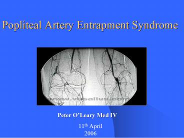
Popliteal Artery Entrapment Syndrome
Peter OLeary Med IV
11th April 2006
2
Aims
- Background of PAES
- Review anatomy of popliteal fossa
- Classify PAES
- Look at a case
- PC
- Exam
- Investigations
- Differential Dx
- Surgical Treatment
- Outcome
3
Introduction
- Popliteal Artery Entrapment Syndrome (PAES)
- Popliteal entrapment was first described by a
Scottish medical student, T. P. Anderson Stuart,
in 1879 - Crush syndrome resulting from compression of the
popliteal artery and impairment of its blood flow
by structures of the popliteal space - Non atheromatous cause of acute limb ischaemia
- Early diagnosis is important
- Very disabling
4
Epidemiology
- 3-5 with anatomic predisposition (in pm exams of
patients with symptomatic vasc disease) - Males 81 Females
- Precise incidence rate is unknown (400 cases in
literature) - 2 types Congenital form/functional form
- Young people (80 personnel
- 25 bilateral
5
?Tibial nerve
Adductor hiatus ?
Popliteal Artery ?
?Popliteal Vein
- Popliteal Artery
- Popliteal Vein
- Tibial Nerve
- Gastrocnemius Lat and Med
- Adductor hiatus
Lateral head of gastrocnemius
Medial Head of gastrocnemius
6
Classification Type I
- Popliteal artery passes medial to and under a
normal medial gastrocnemius head - The vein remains in its normal position
7
Type II
- Medial head of the gastroc inserts more lateral
than normal - Artery descends in a straighter path around the
medial margin of the muscle
8
Type III
- Artery is compressed by a slip of the medial head
arising more laterally than normal - Artery passes through the body of the medial head
in a relatively straight path
9
Type IV
- Popliteal artery passes deep to the popliteus
muscle or a band, with or without associated
gastroc abnormality - Reflects a persistence of a more primitive
embryological vascular pattern of the leg
10
Type VI functional
- Extrinsic compression of the popliteal artery
without identification of anatomical alterations
- Hypertrophy of the gastrocnemius muscle
11
(No Transcript)
12
Presenting Complaint
- 21 yo male
- Sharp pain in right and left calf and great toe
- Began 2 years ago and was becoming progressively
more debilitating - Pain was exacerbated by walking, not running
- Alleviated with rest
- Feet became cold and pale and he experienced
numbness in the digits - Toe and calf pain became severe if he stood for
10 or more minutes
13
Examination
- Unremarkable except
- Active Plantar flexion and passive dorsiflexion
of the foot caused diminished dorsalis pedis and
post tibial pulses - Patient also had a pigeon toed walking gait
foot turned inwards
14
6 Ps Acute limb ischaemia
- Pulselessness
- Pain
- Pallor
- Poikilothermy (cold)
- Paresthesia
- Paralysis
15
Investigations
- Investigations used to diagnose popliteal artery
entrapment and to grade the severity of
circulatory insufficiency and arterial damage - Duplex doppler ultrasound screening exam
- Digital Subtraction Angiography (Gold Standard)
- CT scan
16
(No Transcript)
17
Duplex Doppler ultrasound
- Knee in normal position and doppler shows no
abnormality - Plantar extension of foot and doppler reveals
highly phasic, staccato waveforms, which suggest
high-grade distal arterial stenosis - Peak systolic and end-diastolic velocities can be
measured
18
Ankle-Brachial Index (ABI)
.
- Ratio of the higher systolic blood pressures
between the dorsalis pedis and the posterior
tibial artery to the higher of the systolic blood
pressures in the two brachial arteries - ABI values relate to severity of PAD
19
CT scan
- Submaximal calf muscle contraction demonstrates
accessory head of the gastrocnemius muscle (white
arrow) compressing small-caliber right popliteal
artery (black arrow) - In comparison with the opposite normal-caliber
left popliteal artery (arrow)
20
Intra-arterial Digital Subtraction Angiography
- Images are acquired in digital format
- Blood vessels can be shown in a
near-instantaneous film - Right transfemoral digital subtraction angiogram
shows normal popliteal artery flow - Knee in neutral position
21
Digital Subtraction Angiography
- Severe stenosis of the popliteal artery (Black
arrow) - Full extension at the knee and active plantar
extension at the ankle
22
Differential Diagnosis
23
Histologic changes with continued entrapment
24
Treatment
- The principles surrounding treatment of PAES are
release of the arterial compression, restoration
of as near-normal anatomy as possible, and
preservation or restoration of arterial flow to
the limb - Surgery is the mandatory treatment of PAES except
in the case of asymptomatic functional PAES - If fibrosis has reached a point where there is
aneurysm formation and thrombogenic activity in
the artery simple release of the artery will not
prevent worsening of the situation - Therefore vascular reconstruction may be
necessary
25
Surgical Treatment
- Diagnosis and surgery at an early stage will
decrease the need for vascular reconstruction - A simple myotomy is all that is required
- This will result in fewer complications and a
better prognosis - Musculotendinous section in the absence of
arterial damage is the procedure carried out on
this 21 year old male
26
Musculotendinous section in the absence of
arterial damage
- A posterior approach, via an S- or Z-shaped
incision allows - Greater flexibility for the surgeon
- Closer inspection of the structures within the
popliteal fossa - Sufficient exposure for arterial reconstruction
if required - Allows short saphanous vein to be harvested
27
- The major obstruction was at the medial head of
the gastrocnemius tendon - Tendon was incised
- And removed from around the popliteal artery
- Popliteal artery was followed to the adductor
hiatus - Found to be free of obstruction
28
Complications
- Symptoms, such as claudication, may occasionally
persist or recur postoperatively. - If this occurs in the early postoperative period
then incomplete division of the aberrant
musculotendinous portion must be considered. - In this event, further operative intervention may
be required. - Other complications may occur as a result of
operative intervention such as infection and
hematoma formation.
29
Other therapies
- Endoluminal procedures including intra-arterial
thrombolysis, percutaneous transluminal
thromboembolectomy, and percutaneous transluminal
dilatation are a possibility but they will not
prevent ongoing problems in the presence of
fibrosis and vascular reconstruction is required - Thrombolytic therapy to improve distal runoff is
described as useful to its use in patients with
thrombosed popliteal artery aneurysms. This
appears a reasonable assertion and may reduce the
extent of, if not the need for, reconstruction.
30
Prognosis
- The most exhaustive follow-up study was performed
by di Marzo et al. They reported both a
statistically significant lower complication rate
for MTS as well as a higher patency rate with MTS
(95) over reconstruction (65). - Also, patients undergoing MTS were better able to
undertake a standardized treadmill test than
those following reconstruction (96 vs. 67). - These results may be explained, in part, by the
significantly worse runoff status (P and increased likelihood of presence of
thrombosis, aneurysm, or both (P patients who subsequently underwent
reconstruction.
31
Outcome
- Patient was discharged following surgery
- Returned to work within three weeks of surgery
- Completely asymptomatic following wound healing
- At last follow-up could stand for prolonged
periods without calf or foot pain
32
References
- Principles and Practice of Surgery 4th ed by J.
Garden, A.W. Bradbury, J. Forsythe - Essentials of General Surgery 3rd ed by
P.Lawrence - Ind J Radiol Imag 2002 12191-93
- J Vasc Br 20032(3)210-8
- Di Marzo L, et al. Popliteal artery entrapment
syndrome the role of early diagnosis and
treatment. Surgery 1997, 12226-31 - Popliteal Artery Entrapment Syndrome Mark F.
Henry, MRCS, Denis C. Wilkins, MS, FRCS, and
Anthony W. Lambert, MS, FRCS (Gen Surg) Current
Treatment Options in Cardiovascular Medicine
2004, 6113-120
33
(No Transcript)
Recommended
CrystalGraphics Presentations


