
Circulation - PowerPoint PPT Presentation
1 / 42
Title:
Circulation
Description:
White blood cells spend most of their time outside the circulatory system, ... Shier,David, Jackie Butler, Ricki Lewis: Hole's Human Anatomy and Physiology ... – PowerPoint PPT presentation
Number of Views:114
Avg rating:3.0/5.0
Title: Circulation
1
Circulation Blood
2
Introduction
- organism must exchange materials and energy with
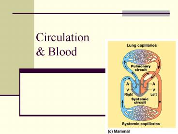
its environment, and this exchange ultimately
occurs at the cellular level - resources that they need, such as nutrients and
oxygen, move across the plasma membrane to the
cytoplasm.Metabolic wastes, such as carbon
dioxide, move out of the cell. - Most animals have organ systems specialized for
exchanging materials with the environment, but
the methods vary.
3
Cont
- bulk transport of fluids throughout the body
connects the aqueous environment of the body
cells to the organs that exchange gases, absorb
nutrients, and dispose of wastes. - Ex, in the mammalian lung, oxygen from inhaled
air diffuses across a thin epithelium and into
the blood, while carbon dioxide diffuses out. - Bulk fluid movement in the circulatory system,
powered by the heart, quickly carries the
oxygen-rich blood to all parts of the body. - As the blood streams through the tissues within
microscopic vessels called capillaries, chemicals
are transported between blood and the
interstitial fluid that bathes the cells.
4
Open Circulation
- In insects, other arthropods, and most mollusks,
blood bathes organs directly in an open
circulatory system. - There is no distinction between blood and
interstitial fluid, collectively called
hemolymph. - One or more hearts pump the hemolymph into
interconnected sinuses surrounding the organs,
allowing exchange between hemolymph and body
cells.
5
Closed Circulation
- closed circulatory system, as found in
earthworms, squid, octopuses, and vertebrates,
blood is confined to vessels and is distinct from
the interstitial fluid. - One or more hearts pump blood into large vessels
that branch into smaller ones cursing through
organs. - Materials are exchanged by diffusion between the
blood and the interstitial fluid bathing the
cells.
6
Quest For Perfection
- closed circulatory system of humans and other
vertebrates is often called the cardiovascular
system. - The heart consists of one atrium or two atria,
the chambers that receive blood returning to the
heart, and one or two ventricles, the chambers
that pump blood out of the heart - Arteries, veins, and capillaries are the three
main kinds of blood vessels. - Arteries and veins are distinguished by the
direction in which they carry blood, not by the
characteristics of the blood they carry. - All arteries carry blood from the heart toward
capillaries. - Veins return blood to the heart from capillaries
7
The Roadways
- Arteries carry blood away from the heart to
organs. - Within organs, arteries branch into arterioles,
small vessels that convey blood to capillaries. - Capillaries with very thin, porous walls form
networks, called capillary beds, that infiltrate
each tissue. - Chemicals, including dissolved gases, are
exchanged across the thin walls of the
capillaries between the blood and interstitial
fluid. - capillaries converge into venules, and venules
converge into veins, which return blood to the
heart.
8
General Rules
- animals with high metabolic rates, mammals, have
more complex circulatory systems and more
powerful hearts than animals with low metabolic
rates, reptiles. - Similarly, the complexity and number of blood
vessels in a particular organ are correlated with
that organs metabolic requirements - Ex. Heart has more arteries and veins then the
bicep.
9
Fish2 Chambers
10
Amphibians and Reptiles
Have 3 chambered heart not so efficient since it
allows oxygenated and unoxygenated blood to mix.
Also have double circulation. Reptiles more
advanced-their heart is almost divided into 4
chambers.
11
Best of the Best
- crocodilians, birds, and mammals, the ventricle
is completely divided into separate right and
left chambers. - left side of the heart receives and pumps only
oxygen-rich blood, while the right side handles
only oxygen-poor blood. - prevents mixing of oxygen-rich and oxygen-poor
blood.
12
Evolution
- evolution of a powerful four-chambered heart was
an essential adaptation in support of the
endothermic way of life characteristic of birds
and mammals. - Endotherms use about ten times as much energy as
ectotherms of the same size. - Therefore, the endotherm circulatory system needs
to deliver about ten times as much fuel and O2 to
their tissues and remove ten times as much wastes
and CO2
13
Circulation
14
(1) The right ventricle pumps blood to the lungs
via (2) the pulmonary arteries. As blood flows
through (3) capillary beds in the right and left
lungs, it loads O2 and unloads CO2. Oxygen-rich
blood returns from the lungs via the pulmonary
veins to (4) the left atrium of the heart. Next,
the oxygen-rich blood blows to (5) the left
ventricle, as the ventricle opens and the atrium
contracts. The left ventricle pumps oxygen-rich
blood out to the body tissues through the
systemic circulation. Blood leaves the left
ventricle via (6) the aorta, which conveys blood
to arteries leading throughout the body. The
first branches from the aorta are the coronary
arteries, which supply blood to the heart
muscle. The next branches lead to capillary beds
(7) in the head and arms.
15
- The aorta continues in a posterior direction,
supplying oxygen-rich blood to arteries leading
to (8) arterioles and capillary beds in the
abdominal organs and legs. - Within the capillaries, blood gives up much of
its O2 and picks up CO2 produced by cellular
respiration. - Venous return to the right side of the heart
begins as capillaries rejoin to form venules and
then veins. - Oxygen-poor blood from the head, neck, and
forelimbs is channeled into a large vein called
(9) the anterior (or superior) vena cava. - Another large vein called the (10) posterior (or
inferior) vena cava drains blood from the trunk
and hind limbs. - The two venae cavae empty their blood into (11)
the right atrium, from which the oxygen-poor
blood flows into the right ventricle.
16
(No Transcript)
17
Cardiac Cycle
- cardiac cycle is one complete sequence of
pumping, as the heart contracts, and filling, as
it relaxes and its chambers fill with blood. - The contraction phase is called systole, and the
relaxation phase is called diastole. - human at rest with a pulse of about 75 beat per
minute, one complete cardiac cycle takes about
0.8 sec. - (1) During the relaxation phase (atria and
ventricles in diastole) lasting about 0.4 sec,
blood returning from the large veins flows into
atria and ventricles. - (2) A brief period (about 0.1 sec) of atrial
systole forces all the remaining blood out of the
atria and into the ventricles. - (3) During the remaining 0.3 sec of the cycle,
ventricular systole pumps blood into the large
arteries.
18
Cardiac Output
- output depends on two factors the rate of
contraction or heart rate (number of beats per
second) and stroke volume, the amount of blood
pumped by the left ventricle in each contraction - average stroke volume for a human is about 75 mL.
- The typical resting cardiac output, about 5.25 L
/ min, is about equivalent to the total volume of
blood in the human body. - Cardiac output can increase about fivefold during
heavy exercise. - Avg. Human Heart Beats 3 mill/year 2.9 mill
liters of blood or 765,000 gallons
19
Cardiac cycle is regulated by electrical impulses
20
Arteries and Veins Differ
- built of similar tissues.
- The walls of both arteries and veins have three
similar layers. - On the outside, a layer of connective tissue with
elastic fibers allows the vessel to stretch and
recoil. - A middle layer has smooth muscle and more elastic
fibers. - Lining the lumen of all blood vessels, including
capillaries, is an endothelium, a single layer of
flattened cells that minimizes resistance to
blood flow.
21
Cont
- differences correlate with the different
functions of arteries, veins, and capillaries. - Capillaries lack the two outer layers and their
very thin walls consist of only endothelium and
its basement membrane, thus enhancing exchange. - Arteries have thicker middle and outer layers
than veins. - The thicker walls of arteries provide strength to
accommodate blood pumped rapidly and at high
pressure by the heart - thinner-walled veins convey blood back to the
heart at low velocity and pressure. - Blood flows mostly as a result of skeletal muscle
contractions when we move that squeeze blood in
veins
22
(No Transcript)
23
Veins have valves to prevent back flow
24
Transfer----The Exchange of Nutrients and Gases
- Occurs at the capillary level. At any given time,
only about 5-10 of the bodys capillaries have
blood flowing through them. - Capillaries in the brain, heart, kidneys, and
liver are usually filled to capacity, but in many
other sites, the blood supply varies over times
as blood is diverted. - For example, after a meal blood supply to the
digestive tract increases. - During strenuous exercise, blood is diverted from
the digestive tract and supplied to skeletal
muscles
25
How to Control Blood Flow
- 2 mechanisms, both dependent on smooth muscles
controlled by nerve signals and hormones,
regulate the distribution of blood in capillary
beds. - 1 mechanism, contraction of the smooth muscle
layer in the wall of an arteriole constricts the
vessel, decreasing blood flow through it to a
capillary bed. - When the muscle layer relaxes, the arteriole
dilates, allowing blood to enter the capillaries. - The other mechanism, rings of smooth muscles,
called precapillary sphincters because they are
located at the entrance to capillary beds,
control the flow of blood between arterioles and
venules
26
Control is GOOD
27
Exchange
- exchange of substances between the blood and the
interstitial fluid that bathes the cells takes
place across the thin endothelial walls of the
capillaries - some substances are carried across endothelial
cells in vesicles formed by endocytosis on one
side and then release their contents by
exocytosis on the other side. - Others simply diffuse between the blood and the
interstitial fluid
28
Bulk Flow (Very Important)
29
Fluid Filtration
- Fluids and some blood proteins that leak from the
capillaries into the interstitial fluid are
returned to the blood via the lymphatic system. - tiny lymph capillaries intermingled among
capillaries of the cardiovascular system - the fluid is called lymph, with a composition
similar to the interstitial fluid - drains into the circulatory system near the
junction of the venae cavae with the right atrium - Along a lymph vessels are organs called lymph
nodes. - The lymph nodes filter the lymph and attack
viruses and bacteria.
30
Blood- The Giver of Life
- invertebrates with open circulation, blood
(hemolymph) is not different from interstitial
fluid. - However, blood in the closed circulatory systems
of vertebrates is a specialized connective tissue
consisting of several kinds of cells suspended in
a liquid matrix called plasma. - plasma includes the cellular elements (cells and
cell fragments), which occupy about 45 of the
blood volume, and the transparent, straw-colored
plasma 55 - plasma consists of water, ions, plasma proteins,
nutrients, waste products, respiratory gases, and
hormones, while the cellular elements include red
and white blood cells and platelets
31
(No Transcript)
32
Blood Cells
- 2 classes of cells red blood cells which
transport oxygen, and white blood cells, which
function in defense. - 3 cellular element, platelets, are pieces of
cells that are involved in clotting - erythrocytes, are by far the most numerous blood
cells. 25 trillion red cells in the bodys 5 L of
blood. - function of red blood cells, oxygen transport
- erythrocytes lack nuclei, an unusual
characteristic that leaves more space in the tiny
cells for hemoglobin, the iron-containing protein
that transports oxygen - RBCs also lack mitochondria and generate their
ATP exclusively by anaerobic metabolism
33
Cont
- An erythrocyte contains about 250 million
molecules of hemoglobin. - Each hemoglobin molecule binds up to four
molecules of O2. WOW!!!!!! - Recent research has also found that hemoglobin
also binds the gaseous molecule nitric oxide (NO) - In the systemic capillaries, hemoglobin unloads
oxygen and it then diffuses into body cells. - The NO relaxes the capillary walls, allowing them
to expand, helping delivery of O2 to the cells - 5 major types of white blood cells, or
leukocytes monocytes, neutrophils, basophils,
eosinophils, and lymphocytes
34
Defense---Defense
- function is to fight infection.
- EX, monocytes and neutrophils are phagocytes,
which engulf and digest bacteria and debris from
our own cells - Lymphocytes develop into specialized B cells and
T cells, which produce the immune response
against foreign substances - White blood cells spend most of their time
outside the circulatory system, patrolling
through interstitial fluid and the lymphatic
system, fighting pathogens
35
Nothing Lasts Forever
- cellular elements of blood wear out and are
replaced constantly throughout a persons life. - For example, erythrocytes usually circulate for
only about 3 to 4 months and are then destroyed
by phagocytic cells in the liver and spleen. - Enzymes digest the old cells macromolecules, and
the monomers are recycled.
36
(No Transcript)
37
Control of RBC Production
- If the tissues do not produce enough oxygen, the
kidney converts a plasma protein to a hormone
called erythropoietin, which stimulates
production of erythrocytes. - If blood is delivering more oxygen than the
tissues can use, the level of erythropoietin is
reduced, and erythrocyte production slows.
38
Making RBCs
39
Clot Formation
40
Major Arteries
41
Major Veins
42
References
- Jack Brown M.S. Biology
- Shier,David, Jackie Butler, Ricki Lewis Holes
Human Anatomy and Physiology 10th edition 2004
McGraw-Hill - Marieb, Elaine Essentials of Human Anatomy and
Physiology 7th edition. 2003 Pearson Education
Inc Benjamin Cummings pub. - Microsoft Encarta Encyclopedia 2004
- Starr and Taggart The Unity and Diversity of
Life 10th edition 2004 Thomson Brookes/Cole - Campbell and Reece Biology 6th edition 2002
Benjamin Cummings. - Raven and Johnson Holt Biology 2004 Holt,
Rinehart and Winston.
Recommended
CrystalGraphics Presentations


