
HERPETOVIRIDAE - PowerPoint PPT Presentation
1 / 51
Title: HERPETOVIRIDAE
1
HERPETOVIRIDAE
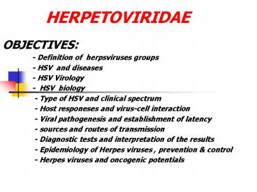
- OBJECTIVES
- - Definition of herpsviruses
groups - - HSV and diseases
- - HSV Virology
- - HSV biology
- - Type of HSV and clinical
spectrum - - Host responeses and virus-cell
interaction - - Viral pathogenesis and
establishment of latency - - sources and routes of
transmission - - Diagnostic tests and
interpretation of the results - - Epidemiology of Herpes viruses ,
prevention control - - Herpes viruses and oncogenic
potentials
2
Herpesviridae
- Large family of large, complex viruses
- Infect vertebrate hosts
- Three subfamilies
- Alphaherpesvirinae, 2 genera
- Betaherpesvirinae, 3 genera
- Gammaherpesvirinae, 2 genera
- Very important as human pathogens
- Cause cold sores, genital herpes, chicken pox,
shingles, mononucleosis and many other diseases - Infection is for life herpesviruses become
latent in hosts, then reactivate
3
Herpesviruses
- HHV1, Herpes simplex 1
- Cold sores, epithelial and neuronal cells
- HHV2, Herpes simplex 2
- Sexually transmitted disease (STD), also as
above, teratogenic, can be fatal in newborns - HHV3, Varicella-zoster
- Chicken pox, shingles
- HHV4, Epstein Barr virus
- Mononucleosis, Burkitts lymphoma, lymphoid
tissue only - HHV5, Cytomegalovirus
- Salivary gland tropic, teratogenic, can be fatal
in newborns - HHV6, Roseolovirus
- Childhood rash, multiple sclerosis?
- HHV8
- Associated with Kaposis sarcoma
HSV 6,7,8 all identified after 1990, after HIV
4
Cryo-EM shows regular, external structure
preserved
Irregular structures often seen in micrographs
are artifacts of distortion from negative staining
Nucleocapsid contains core of protein wrapped in
genomic DNA
5
Herpesvirus particles
- Genome Single large segment of dsDNA, 3 of
particle weight (124-235 kbp) - Core Nucleic acid wrapped around cylindrical
structure 25-30 nm - Capsid T16 icosahedron composed of 162
capsomeres (150 hexamers and 12 pentamers),
capsid diameter 100-110 - Tegument poorly defined material between capsid
and envelope, contains alpha trans-inducing
factor (a-TIP) necessary for activation of a
genes and virion host shutoff protein (VHS) - Envelope Derived from nuclear membrane,
surrounds tegument, has spike glycoproteins
virion diameter 120-200 nm
6
Human Herpesvirus 1 particles and genome
organization
7
Biology of herpesviruses
- All specify a large array of enzymes involved in
nucleic acid metabolism - Virus DNA synthesis and capsid assembly in
nucleus - Production of infectious progeny is accompanied
by destruction of infected cell - A single virus can cause several diseases
- Herpesviruses remain latent in the host for life,
and can be reactivated to cause lesions at or
near the initial infection site
8
HSV Establishes Latent Infections
- Once infection has taken place HSV can remain
dormant for months, years, lifetime - Cell types that HSV can infect and remain dormant
- Neurons, B-cells and T-cells
- Examples
- Shingles which can appear years after first
chickepox infection (caused by varicella zoster,
causes both chickenpox and shingles) - Genital Herpes outbreaks
9
Virus Latency
10
Types of HSV
- The Type 1 virus causes cold sores. Most people
get Type 1 infections during infancy or
childhood.
- The Type 2 virus causes genital sores. Most
people get Type 2 infections following sexual
contact with an infected person. ii
Both types can be differentiated by biologic,
biochemical and antigenic properties
11
Symptoms
- Mouth sores
- Genital lesions (male) -- may be preceded by
burning or tingling sensation - Genital lesions (female) -- may be preceded by
burning or tingling sensation - Blisters and/or ulcers -- most frequent on the
mouth, lips and gums or genitalia - Fever blisters
- Fever -- may be present especially during the
first episode - Enlargement of lymph nodes in the neck or groin
12
Immunology
The constant battle between our body and invaders
- T cells can prevent HSV-1 reactivation from
latency in sensory neurons with the help of gamma
interferon
- we can produce two different antibodies, one
against each type of HSV
- When both HSV 1 antibodies and HSV 2 antibodies
are present, they can crosslink with one another
and neither Antibody will be effective.
- HSV proteins inhibit cellular DNA synthesis and
the virus uses its own primase and other
replication machinery to replicate its DNA.
- .
13
Viral Genes Block Immune Response
- Out of 84 genes only 37 involved in replication
- Some of the remainder involved in blocking immune
response against virus - Vhs and ICP27 block interferon effects by
degrading cellular mRNAs - ICP47 binds transporter proteins that aid antigen
presentation - Self and viral peptides are constantly being
presented thru MHC I and provoke immune responses
when appropriate - ICP47 prevents transport of viral peptides on
surface of cell - ? no viral antigen presentation which means no
immune response
14
HSV TYPE-SPECIFIC SEROLOGY
- Subclinical genital herpes in the mother in late
pregnancy is responsible for most neonatal herpes
cases. - HSV type-specific serology should be used for
prenatal testing. - Counseling safe sexual practices, abstinence
should be provided to patient/partner, depending
on results of the serologic testing.
15
Diagnosis of HSV
- The appearance of HSV is often so typical that no
testing is needed to confirm an infection, only a
physical. - The genital herpes sores may not be visible to
the naked eye so a doctor may have to do a swab
from the infected skin (culture) and send it to
the lab for analysis. - A viral PCR can be run on a swab of infected
tissue. - A blood test, can show if a person has been
infected with HSV but cannot distinguish between
type I and II. - There are also newer blood tests that can tell
whether a person has been infected with HSV 1
and/or 2 by the patients immune response to the
herpes infection.
16
HSV DIAGNOSTIC TESTS
- Tzanck prep low sensitivity, not recommended
as diagnostic test - DFA stain scrapings same day, sensitive
- ELISA detection of HSV in skin lesions
- Eye consult
- EEG, MRI (temporal lobe involved)
- Histopathology
17
HSV-CULTURES
- Culture at 24 - 48 hrs results in 2-7dskin,
conjunctiva, mouth/nasopharynx, rectal, urine,
blood, CSF - Hi-risk /symptomatic Rx pending cultures
- Positive cultures from any site obtained gt 48hrs
viral replication rather than colonization - CSF cultures usually negative in encephalitis
- PCR DNA-CSF test of choice test before
after RX
18
CMV DNA PCR CMV-IgM
- CMV DNA PCR
- CSF preferred test
- positive result poor neurologic outcome.
- Blood positive active infection associated
with hearing loss - Newborn heel stick dried blood spots
opportunity for universal screening? - CMV IgM not recommended for neonates
- False-positives and false-negatives occur
19
CHILD CARE CENTERS
- Plastic surfaces and toys harbor CMV for hours
- Viruria 70 infected gt18 mth old children.
- 30 seroconversion among mothers whose children
shed CMV vs 0 if children dont shed - Child to mother transmission confirmed.
- Young children in child care centers are
important source of primary CMV infection for
pregnant women.
20
MATERNAL DIAGNOSIS Is it primary infection?
- IgG seroconversion ELISA
- New CMV specific IgM immunoblot
- IgG avidity index Anti CMV IgG has low avidity
for first 14 wks after conversion - IgG avidity index IgMimmunoblot combination
may help identify primary inf.Guerra et al AM.
J Ob Gyn 2000
21
CMV - PRENATAL DIAGNOSISFetus infected?
Symptomatic?
- Viral culture amniotic Fluid
- Viral culture fetal blood
- DNA-PCR quantitative PCR
- CMV-IgM in cordocentesis not very sensitive
- Ultrasound
- Hematologic tests
- Guerra et al AM. J Ob Gyn 2000
22
DAIGNOSIS - CMV CULTURE
- Virus must be isolated in urine /saliva lt3 wks
of age to prove congenital infection - If gt3 wks of age, cannot differentiate
congenital, natal, or postnatal infection unless
the infant previously has had a negative culture.
This distinction is important because congenital
infection is associated with hearing impairment. - Shell-vial culture-helps rapid identification.
23
TRANSFUSION-ACQUIRED CMV
- Characterized by a gray ashen pallor, respiratory
distress, pneumonia, hepatosplenomegaly,
hepatitis, atypical lymphocytosis,
thrombocytopenia, and hemolytic anemia - Infection in LBW infants may be severe
- 10 mortality.
24
BREAST MILK TRANSMISSION
- Seropositive mothers shed virus 20 70 of
the time in BM - 60 term infants fed virus positive breast milk
develop inf but it is benign due to mat(Ig)G. - 15-25 preterm infants (no mat IgG) develop
symptoms, hepatosplenomegaly, pallor,
neutropenia, thrombocytopenia, elevated LFT.
Neurologic sequelae ?? - Co-infection with HIV - risk fctor
25
TRANSPLACENTAL TRANSMISSION
- PRIMARY MATERNAL INFECTION
- 2-6 mothers/yr seroconvert in pregnancy
- Transmission 40 50
- Earlier mat. inf. more severe the fetal inf.
- RECURRENT MATERNAL INFECTIONTransmission
0.5-1.5most infants asymptomatic - REINFECTION new CMV strain
26
EPIDEMIOLOGY
- In developing countries, almost 100.
- After an active replication stage, CMV enters a
latent stage in leukocytes and other tissues.
Like other herpes viruses, CMV reactivates during
relative immuno-compromise, such as pregnancy
27
CONGENITAL CMV INFECTION
- Most common congenital infection 1 newborns
infected 40,000 /yr in US - Leading infectious cause of
- Mental Retardation, Cerebral Palsy, or,
most commonly, hearing impairment - involving gt 8,000 infants/ yr in the US
28
PREVENTION
- Transfusion-acquired infectionCMV
antibody-negative blood products, leukofiltration
of blood to remove WBC,frozen deglycerolized
RBCs as they lack viable leukocytes. - Breast Milk acquired CMVFreezing human milk
20C for 3-7d. Pasteurization (62.5C) not
routinely available. - CMV vaccine - investigational.
29
PREVENTION
- Meticulous hand hygiene after exposure to urine
or saliva from infants and toddlers and
immunocompromised patients - Standard precautions only.
- Routine screening is not recommended for women of
childbearing age as no interventions are
available. - Pregnant women are not excluded from caring for
infants who are infected with CMV.
30
Treatment of HSV
- The combined effects of acyclovir and human
interferon-alpha as drug therapy are being
investigated now. - Hsv 1 protective surface glycoprotein gD
expressed in CHOs and protected mice - To keep from spreading the infection
- Keep the infected area clean and dry to prevent
other infections from developing. - Try and avoid touching the sores.
- Wash your hands after contact with the sores.
- Avoid sexual contact from the time you first feel
any symptoms until the sores are completely
healed.
31
VARICELLA
- Chickenpox in pregnancy rare 5 in 10,000
- Maternal Inf lt 20 weeks gest
- Varicella embryopathy in 2 infants
- Maternal Inf gt 20 weeks gest
- Inapparent varicella, Zoster in early childhood
- Maternal Inf 5d before to 2d after delivery
- Neonatal VZV inf/ pneumonia (can be fatal)
32
HORIZONTAL TRANSMISSION-VARICELLA
- Unusuall
- Most neonates protected by mat. antibody
- 30 wk, lt 1 kg. neonates could be susceptible -
VZIG - Screen rapidly for VZV antibodyVZIG to
susceptible neonates - VZIG to all neonates following exposure ?
33
VARICELLA IN PREGNANCY
Maternal Inf Potential consequences
lt20 wks gest Spont abortion Fetal Varicella Syn - 2
any stage Fetal death, herpes zoster 1st yr of life
Near term5dltdelivery 2dgtdelivery Cong disseminated varicella Varicella pneumonia (can be fatal)
34
EBV Viral Structure
- A core containing a linear, dsDNA molecule of
about 175 kbp. - An icosahedral capsid, approximately 100-110 nm
in diameter, containing 162 capsomeres with a
hole running down the long axis. - An amorphous, sometimes asymmetric material that
surrounds the capsid, designated as the tegument - An envelope containing viral glycoprotein spikes
on its surface.
35
Infection and Replication
36
Replication
- EBV replicates in the epithelial cells of the
mouth, tongue, salivary glands, and oral cavity
(few symptoms may be present, but a person can
still be infectious) - EBV infects B cells in the lymph nodes of the
oral cavity - Once inside B cells, EBV expresses proteins
- Nucleus EBNA (Epstein-Barr Virus Nuclear
Antigens) - Plasma Membrane LMP (Latent Membrane Proteins)
- Expression of these proteins stimulates B cell
replication in lymph nodes producing clones - Since many B cells are infected, polyclonal
B-cell growth occurs which allows the disease to
begin a long time after initial exposure to EBV
37
Site of Infection
- Infection of Epithelial Cells by EBV in vitro
- Active replication, production of virus, lysis of
cell - Infection of B cells by EBV in vitro
- Latent infection, with immortalization
(proliferate indefinitely) of the virus-infected
B cells - Linear EBV genome becomes circular, forming an
episome, and the genome usually remains latent in
these B cells - Viral replication is spontaneously activated in
only a small percentage of latently infected B
cells. - Signal transduction pathways can reactivate EBV
from the latent state
38
Primary Infection Diseases
- Infectious Mononucleosis (glandular fever) -
fever, lymphadenopathy, and pharyngitis
- Chronic active EBV infection - severe illness of
more than six months, histologic evidence of
organ disease, and demonstration of EBV antigens
or EBV DNA in tissue (mimics chronic fatigue
syndrome)
- X-Linked Lymphoproliferative Disease - inherited
disease of males, absence of functional SAP gene
impairs the normal interaction of T and B cells
resulting in unregulated growth of EBV-infected B
cells.
39
Symptoms
- The classic triad of mononucleosis is
- Inflammation of the pharynx (or tonsils) --
usually the severest symptom - Fever (higher in the evening)
- Lymphadenopathy (usually in the neck, groin or
under the arms)
40
Symptoms
- Other symptoms include
- Fatigue and malaise
- Rash (associated with the use of ampicillin)
- Headache
- Muscle aches
- Abdominal pain
- Occasional jaundice
- Enlargement of the spleen and liver
41
Diagnosis of EBV
- Clinical diagnosis- Classic triad of symptoms
lasting 1-4 weeks - Serologic test- Shows elevate white blood cell
count, an increased number of lymphocytes,
greater than 10 atypical lymphocytes, and a
positive reaction to a mono spot test - Someone who appears to have infectious mono, a
positive Paul-Bunnell heterophile antibody test
can be done - Serologic testing is the method of choice for
primary infection - EBV specific lab tests can be performed, testing
patient for EBV antibodies.
42
Immune System to the Rescue! (or not)
- Epithelial cells and polyclonal B cells express
the viral-encoded LMP glycoprotein in their
plasma membranes - Killer T cells recognize the LMP glycoprotein and
kill the EBV-infected cells - While T cells are mounting an attack on B cells,
the immune response of a person is abnormal
producing atypical T cells and antibodies that
can confirm diagnosis of infectious mononucleosis
43
Treatment of EBV
- Infectious Mononucleosis
- No specific therapy just nonaspirins and rest
- Oral Hairy Leukoplakia
- Acyclovir inhibits EBV replication
- EBV Lymphoproliferative Disease
- reduction in the dose of immunosuppressive
medication - Surgical removal or irradiation of localized
lymphoproliferative lesions
44
Prevention and Vaccines
- EBV lymphoproliferative disease
- infusion of B-celldepleted marrow to offset the
proliferation of donor B cells - Removal of donor B cells along with T cells
- Vaccination studies underway but no vaccine found
so far
45
Cancers Associated with EBV
- Nasopharyngeal Carcinoma
- Southern China, Northern Africa, and Alaskan
Eskimos - Elevated titers of IgA antibody to EBV structural
proteins - Burkitt's Lymphoma
- Found in equitorial Africa and associated with
malaria which doesnt allow T-cells to control
proliferation of EBV-infected B cells - Tumors present in jaw
- Hodgkin's Disease
- EBV DNA detected in tumors
- Lymphoproliferative Disease
- Impaired T-cell immunity and cannot control
proliferation of EBV-infected B cells
46
Complications
- Meningitis-- infection of the sheaths and
membranes (meninges) covering the brain and the
spinal cord. - Encephalitis-- acute inflammation of the brain,
commonly caused by a viral infection by insect
bites or food and drink - Eczema herpetiform-- widespread herpes across the
skin) - Keratoconjunctivitis-- Infection of the eye
- Prolonged, severe infection in immunosuppressed
individuals - Pneumonia
- Infection of the trachea
- Keratitis-- Corneal infection, irritations, and
inflammations
47
CONCEPT QUESTIONS
- -Herpesviruses are the causative agents of
- -Mention the types of HSV and their related
diseases. - - List the structural components their
characteristic - of HSV.
- - Mention the biology of HSV.
- - Define virus latency .
- - Describe latency in HSV
- - Tabulate differences between HSV-1 X HSV-2
- - What are the main syndromes of HSV infections?
- - Describe the HSV gene expression.
- - What are the immunologic pattern of HSV
infection?
48
- - How viral genes blocks host immune responses?
- - What are the uses of HSV type-specific serology
? - - Mention the basis for HSV diagnosis .
- - What are the diagnostic test for HSV.
- - For what purposes HSV culture is required?
- - What are the significance of CMV ?
- - Mention the diagnostic methods for CMV
detection. - - What is the impact of day-care centers on CMV
spread - -Why maternal diagnosis of CMV infection is
important? - - What types of tests used for CMV prenatal
diagnosis ? - - CMV culture is useful in what ?
- - What are the modes of CMV transmission ?
49
- -What are the epidemiological features of CMV?
- - Define congenital CMV infections?
- - What are the preventive measure for CMV
infections? - -Why acyclovir can be considered as anti-herpes ?
- - Define VZV .
- - What is the impact of VZV infections near term?
- - What are the expected outcome of varicella in
pregnancy ? - - Describe the structure of EBV.
- -Describe EBV infection and replication.
- - Mention the pathogenesis of EBV .
- - What is the sites of EBV infections?
- - What are the primary infections of EBV?
- - In what way EBV can evade host immune system?
- - What is the diagnostic test for infectious
mononucleosis? - - What are the preventive measure to EBV
infections? - - Mention the canccers associated with EBV
infections. - - What are the major complications associated
with herpesviruses infections?
50
Family Poxviridae (poxviruses) The family name
is taken from the major disease symptom caused by
these organisms, the pox. The pox is an elevated
lesion of the skin. The members of this family
are the largest of all the viruses and are
considered to be an evolutionary intermediate
between the viruses and the bacteria. The viral
particles (sometimes called elementary bodies)
are somewhat rounded, brick-shaped, or ovoid, and
have a complex structure consisting of an
internal central mass, the nucleoid, surrounded
by two membrane layers. The surface is covered
with ridges which may be tubules or threads. All
poxviruses are related immunologically by a
common internal antigen. They are divided into
genera on the basis of their more specific
antigens, nucleic acid homology, morphology and
natural hosts. The entire replication cycle
occurs in the cytoplasm. The viruses causing
human disease include
Family Poxviridae (poxviruses)
The family name is taken from the major disease
symptom caused by these organisms, the pox. The
pox is an elevated lesion of the skin. The
members of this family are the largest of all the
viruses and are considered to be an evolutionary
intermediate between the viruses and the
bacteria. The viral particles (sometimes called
elementary bodies) are somewhat rounded,
brick-shaped, or ovoid, and have a complex
structure consisting of an internal central mass,
the nucleoid, surrounded by two membrane layers.
The surface is covered with ridges which may be
tubules or threads. All poxviruses are related
immunologically by a common internal antigen.
They are divided into genera on the basis of
their more specific antigens, nucleic acid
homology, morphology and natural hosts. The
entire replication cycle occurs in the cytoplasm.
The viruses causing human Variola virus - the
agent of smallpox, an infection of
reticuloendothelial, vascular endothelial and
epithelial cells Monkeypox virus - the agent of
monkeypox, a disease similar to smallpox but with
the additional symptoms of cervical and inguinal
lymphadenopathy disease include
51
Variola virus - the agent of smallpox, an
infection of reticuloendothelial, vascular
endothelial and epithelial cells Monkeypox virus
- the agent of monkeypox, a disease similar to
smallpox but with the additional symptoms of
cervical and inguinal lymphadenopathy Vaccinia
virus - used to vaccinate against smallpox. It
causes a localized exanthem through epithelial
cell infection. Cowpox virus - the agent of
cowpox, a self-limiting disease resulting in
vesicles and pustules of the hands Orf virus -
the agent of contagious pustular dermatitis, an
epithelial cell infection Pseudocowpox virus -
the agent of pseudocowpox (Milker's nodules,
paravaccinia). This is an epithelial cell
infection. Molluscum contagiosum virus - this
causes molluscum contagiosum, a self-limiting
infection of epithelial cells Yaba monkey tumor
virus - this causes a histiocytoma of the head or
limbs Tanapox virus - causes tanapox, a
self-limiting epithelial cell infection
Recommended
CrystalGraphics Presentations


