
Cell Membrane/Plasma Membrane - PowerPoint PPT Presentation
1 / 19
Title:
Cell Membrane/Plasma Membrane
Description:
Cell Membrane/Plasma Membrane functions: 1. integrity of the cell 2. controls transport = selectively permeable 3. excludes unwanted materials from entering the ... – PowerPoint PPT presentation
Number of Views:64
Avg rating:3.0/5.0
Title: Cell Membrane/Plasma Membrane
1
Cell Membrane/Plasma Membrane
- functions 1. integrity of the cell
- 2. controls transport selectively
permeable - 3. excludes unwanted materials from
entering the cell - 4. maintains the ionic concentration of the
cell osmotic pressure - of the cytosol
- 5. forms contacts with neighbouring cells
tissue
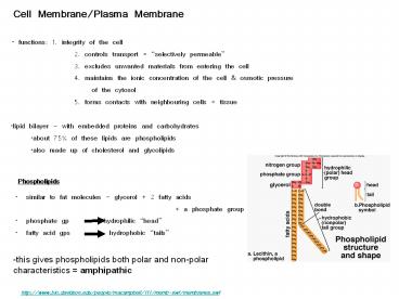
- lipid bilayer - with embedded proteins and
carbohydrates - about 75 of these lipids are phospholipids
- also made up of cholesterol and glycolipids
Phospholipids
-this gives phospholipids both polar and
non-polar characteristics amphipathic
http//www.bio.davidson.edu/people/macampbell/111/
memb-swf/membranes.swf
2
polar heads out
non-polar tails in
-the polar and non-polar attributes of the lipids
results in a bilayer arrangement -cholesterol is
also polar (OH group) and non-polar (steroid
rings) and contributes to this arrangement OH
group faces out and the steroid rings face inward
3
- membrane proteins
4
Functions of Integral Proteins
-in addition 4. enzymes 5. linkers anchor
proteins of the PM to the protein filaments
inside or to neighboring cells 6. cell-identity
markers used in identifying self by the
immune system e.g MHC proteins e.g. ABO blood
typing
5
B. Membrane function
- Physical isolation - from the surrounding ECF
- -allows the cell to create different environments
outside and inside - -allows for the creation of gradients
electrical and chemical
2. Integrity of cell - cell shape and
size -increase cell size, increase surface
area/volume -increase exchange surface
3. Sensitivity - first part of cell that is
affected by changes in the extracellular
environment
4. Structural support - connections between cells
provides tissues with support and stability
5. Controls transport selectively
permeable -two types Passive - Diffusion,
Osmosis, Facilitated Active - Active
transport, Exocytosis, Endocytosis,
6
Membrane Gradients
- selective permeability of the PM allows the cells
to control the concentration of ions within the
cell and outside the cell (in the ECF) - this results in a distinct distribution of
positive and negative ions inside and outside the
cell - typically the inside of the cell is more
negatively charged - this difference in electrical charge between
inside and outside electrical gradient - because it occurs across the PM we call this
difference in charge membrane potential - can be measured with tiny glass electrodes
- varies from cell to cell
- very important in the functioning of neurons and
muscle cells
7
Membrane Permeability and Transport
- permeability property that determines the
effectiveness of the PM as a - barrier
- permeability varies depending on the organization
and characterization of - the membrane lipids and proteins
- transport across the membrane may be passive or
active
passive transport
active transport
diffusion osmosis facilitated
endocytosis (pinocytosis phagocytosis receptor-me
diated) exocytosis
http//programs.northlandcollege.edu/biology/Biolo
gy1111/animations/transport1.html
8
- materials may cross into a cell based on
concentration and size - if they cross from high to low they are
traveling with their concentration - gradient requires no energy (Passive)
- -if they cross against the concentration gradient
requires energy (Active) - small particles may cross through the lipid
bilayer - others may require integral proteins that help
(e.g. channels or pores) - others may enter through the fusion of tiny
vesicles with the PM
9
A. Diffusion movement of materials from high
to low -random movement, no energy needs to be
synthesized -the movement is driven by the
inherent kinetic energy of the particles moving
down their concentration gradient -movement
could be through the bilayer itself or
through channel proteins -three ways to
diffuse 1. through the lipid bilayer lipid
soluble (non-polar), alcohol, gases, ammonia,
fat-soluble vitamins 2. through a channel
charged, small ions (polar) -some channels are
gated open and close 3. facilitated
diffusion larger molecules too big for
channels
10
B. Osmosis
-in osmosis the membrane is permeable to water
and NOT to the solutes -but it is the
concentration of solutes that causes the water to
move -experiment U shaped tube divided by a
membrane permeable to water only -increase the
solute concentration in the right half of the
tube -this increases the pressure caused by the
increase solutes osmotic pressure -therefore
increasing solute concentration increases osmotic
pressure -water will move in to decrease this
OP -OP is important in determining how much
fluid remains in your blood and how much leaves
to surround the cells in your tissues
11
-Osmosis is controlled by tonicity degree to
which a the concentration of a specific solute
surrounding a cell causes water to enter or leave
the cell
hypertonic Sin lt Sout, water exits cell
e.g. isotonic Sin Sout, no water
movement
12
C. Facilitated transport molecules move by a
carrier protein from high to low -binds to
a receptor site on the plasma membrane -transport
ed by the carrier protein -no energy
required -but there is a limit to the amount of
FD cells can undergo and it has to do with the
of carrier proteins on the PM
-molecules that are insoluble, too polar or too
large e.g. glucose amino acids
13
A. Active transport molecules are moved against
the the concentration gradient i.e. from low
to high
-two kinds primary and secondary -primary active
transport -requires a protein carrier and
ATP -carrier is often called a pump e.g.
sodium/potassium pump three Na are pumped out
of a cell and 2 K are pumped into the cell (Na/K
ATPase) -Na binds to the pump, ATP then binds
and gets hydrolyzed, a P group attaches to the
pump and changes its shape expels the Na out of
the cell -K then binds the pump and causes the
release of the P, the pump returns to its
original shape, bringing K into the cell
http//highered.mcgraw-hill.com/sites/0072437316/s
tudent_view0/chapter6/animations.html
14
2. secondary active transport -the energy
stored in a concentration gradient is used to
drive the transport of other materials e.g
Na/Ca antiporter opposite direction for Na and
Ca movement primary transport establishes high
Na outside the cell this concentration
gradient creates potential energy which is
stored by the antiporter pump - as Na leaks
back in this potential energy is converted into
kinetic energy which drive the movement of a Ca
ion against its gradient -some pumps can also
pump two materials in the same direction
symporter e.g. Na/glucose symporter -most of
our cells use the energy created by the Na
gradient to power the movement of other ions
15
B. Exocytosis secretion of a substance outside
the cell -made within the cell, packaged into
transport vesicles-gt fusion with the plasma
membrane and release outside the cell e.g.
nerve cells - neurotransmitter release
http//highered.mcgraw-hill.com/sites/0072437316/s
tudent_view0/chapter6/animations.html
16
C. Endocytosis reverse of exocytosis,
internalization of substances -3 forms 1.
pinocytosis cell drinking
17
2. phagocytosis cell eating
18
3. receptor-mediated internalization of
specific substances -binding of a ligand with
its receptor -gt internalization into the
cell -occurs at specific sites within the PM -gt
clathrin-coated pits -internalization at pits
-gt clathrin-coated vesicle -vesicle fuses with
endosomes - processing
http//sumanasinc.com/webcontent/animations/conten
t/endocytosis.html
19
Medical application
- HIV and receptor-mediated endocytosis
- binding of HIV virus to the CD4 protein on the
surface of T helper cells and macrophages
results in the RME of the HIV virus - the HIV viral particles are made by the host cell
protein synthesis machinery and assembled at the
hosts PM released from the cell exocytosis - the infected T cells are killed leading to low T
cell counts in infected people
Recommended
CrystalGraphics Presentations


