
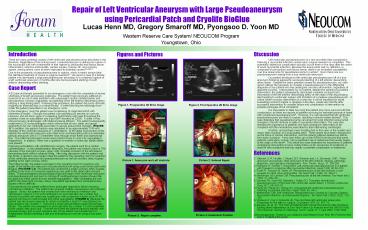
Repair of Left Ventricular Aneurysm with Large - PowerPoint PPT Presentation
1 / 1
Title:
Repair of Left Ventricular Aneurysm with Large
Description:
Repair of Left Ventricular Aneurysm with Large Pseudoaneurysm using Pericardial Patch and Cryolife BioGlue Lucas Henn MD, Gregory Smaroff MD, Pyongsoo D. Yoon MD – PowerPoint PPT presentation
Number of Views:151
Avg rating:3.0/5.0
Title: Repair of Left Ventricular Aneurysm with Large
1
Repair of Left Ventricular Aneurysm with Large
Pseudoaneurysm using Pericardial Patch and
Cryolife BioGlue
Lucas Henn MD, Gregory Smaroff MD, Pyongsoo D.
Yoon MD Western Reserve Care System/ NEOUCOM
Program Youngstown, Ohio
Figures and Pictures
Introduction There are many potential causes of
left ventricular pseudoaneurysms described in the
literature. Regardless of the inciting event, a
pseudoaneurysm is defined as rupture of the
myocardial wall with containment of free rupture
by pericardial and fibrous tissue. Inflammation,
infective endocarditis, cardiac surgery, trauma,
MI, and coronary spasm, among others, have all
been implicated as potential causes2,3,4,5,6,7. Du
e to the propensity of pseudoaneurysms to
rupture, which is nearly always fatal, the
definitive treatment of choice is surgical
resection8. We report a case of a female patient
who developed a large pseudoaneurysm secondary to
a contained rupture of a left ventricular
aneurysm 2 months after she had successful
stenting of a left anterior descending artery
stenting. Case Report A 62-year-old female
presented to our emergency room with the
complaints of severe shortness of breath and
severe weakness. The patient had previously
suffered an acute MI at an out of state
institution with subsequent cardiac
catheterization and percutaneous coronary
angioplasty and stenting of the left anterior
descending artery utilizing a drug eluting stent.
Following this procedure, the patient did poorly
clinically requiring an extended stay in a rehab
facility. A week following her discharge from
rehab the patient presented to our emergency
room. In the emergency room the patient was
complaining of chest discomfort with shortness of
breath. Clinically the patient was saturating 98
percent on room air. However, she did show signs
of congestive heart failure with rales throughout
the posterior chest on auscultation and a pro-BNP
elevated at 22096. A chest X-Ray showed severe
cardiomegaly with bilateral pleural effusion.
The patient subsequently underwent a 2-D
echocardiogram which showed that all mid, distal,
and apical segments to be non contractile
secondary to severe aneurysmal changes. (FIGURE
1) In addition, the ejection fraction was
calculated to be approximately 15 with diameter
of the ventricle measured at 7 centimeters. At
the distal most portion of the septum the
ventricular aneurysm was noted to be
communicating with a 4 centimeter pseudo-aneurysm
with a 1.5 centimeter neck through which there
was bidirectional flow. The mitral and tricuspid
valves were noted to have 3 and 3-4
regurgitation, respectively. In addition, there
was a question of whether a ventricular septal
defect was present. Following consultation with
cardiothoracic surgery, the patient went for a
cardiac catheterization. In the catheterization
laboratory, the patient was noted to have a 70
stenosis of the mid-right coronary artery with
complete occlusion of the left anterior
descending stent without collateral filling.
After long discussion with the patient and her
family, a decision was made to take the patient
to the operating room for resection of the
ventricular aneurysm and pseudoaneurysm as well
as coronary artery bypass grafting to the right
coronary artery. The following day, the patient
was taken to the operating room for resection and
repair of the ventricular aneurysm and
pseudoaneurysm using a pericardial patch and
Bioglue. (PICTURE 1-4) The patient also
underwent a single coronary artery bypass
grafting in the form of a reversed saphenous vein
graft to the distal right coronary artery.
Transesophageal echocardiogram following repair
if the ventricular aneurysm and pseudoaneurysm
and prior to coming off cardiopulmonary bypass
showed the tricuspid and mitral valves to have
minimal regurgitation. After completing the vein
graft to the distal right coronary artery, the
patient separated from cardiopulmonary bypass
without difficulty. Post operatively the
patient suffered from prolonged respiratory
failure requiring mechanical ventilation. The
patient also required multiple vasopressors and
inotropic support. Slowly, the patient was
weaned from the mechanical ventilation and
vasopressor support. A 2-D echocardiogram on post
operative day number four showed concentric left
ventricular hypertrophy with an ejection fraction
to be 55-60 percent with trace to mild tricuspid
and mitral regurgitation. (FIGURE 2) Because the
patient had decreased response to verbal commands
a head CT was obtained on post operative day
number 7. It showed a small right
parietal/occipital hemorrhagic stroke. The
patient was extubated on post operative day
number 12 and transferred to the cardiac step
down unit. Over the next week the patient
improved steadily with physical therapy and on
post operative day 20 the patient was transferred
to a rehabilitation facility tolerating a diet
and ambulating on room air using a four point
walker.
- Discussion
- Left ventricular pseudoaneurysm is a rare and
often fatal complication following a myocardial
infarction unless early surgical resection is
completed. This potentially disastrous
complication typically occurs three or five days
after the onset of acute myocardial infarction.
Because the aneurysmal wall, by definition, is
composed of pericardium and fibrous tissue, there
is an increased susceptibility over true
aneurysms for pseudoaneurysms to rupture1. Even
more rare is a pseudoaneurysm arising from a true
ventricular aneurysm. - Our patient developed a left ventricular
pseudoaneurysm off of a true aneurysm following
apparently successful stenting of a left anterior
descending artery lesion. Despite the many
potential causes for pseudoaneurysm, our case
report outlines the necessity to consider a
pseudoaneurysm in the differential diagnosis of
any patient who has undergone coronary
intervention, regardless of apparent success.
Unfortunately for our patient, despite the
seeming successful intervention, the patient
subsequently developed complete occlusion and
thrombosis of her left anterior descending artery
at the origin of the drug eluding stent leading
to ventricular rupture with formation of a
massive pseudoaneurysm. In a time where coronary
intervention is on the rise, it will be important
for those evaluating recurrent angina or dyspnea
in the days, weeks and months after successful
intervention to consider failure and complication
of intervention as possible etiologies of the
complaints. - It is impossible to state how long this patient
may have survived with this complication, as
there are reports of patients surviving for
extended periods of times with unruptured
pseudoaneurysm6. However, it is well reported
that left ventricular pseudoaneurysms are likely
to rupture, resulting in almost certain demise8.
This case illustrates that pseudoaneurysms can
arise from post infarction aneurysmal changes in
the left ventricle in spite of successful
intervention. The case also stresses the
importance of patients who have undergone
intervention to be compliant with the medications
prescribed by their intervening cardiologist. - A further, and perhaps more troubling fact in
this case, is the sudden and nearly fatal
occlusion of a drug eluting stent. These stents
have been championed as the future of cardiac
intervention, and this patient suffered
deleterious effects from early occlusion. A
two-year follow up on the SIRIUS trial showed
that some patients, particularly women, had a
significant risk of restenosis that usually began
at the origin of the stent9. This article and
case illustrates the importance of patients
undergoing intervention to have routine follow up
for recurrence of symptoms or onset of new
symptoms that may signify a potential
complication or restenosis. - References
- Stewart, S. R. Huddle, I. Stuard, B.F. Shreiner
and J.A. Deweese, 1981. False aneurysm and
pseudo- false aneurysm of the left ventricle
etiology, pathology, diagnosis, and operative
management. Ann. Thorac. Surg., 31 259-265. - 2. Reinecke H, Wichter T, Weyand M. Left
ventricular pseudoaneurysm in a patient with
Dressler's syndrome after myocardial infarction.
Heart 1998 8098100. - 3. Fiorilli R, Tomasco B, Tesler UF.
Pseudoaneurysm of the left ventricle a rare
sequela to mitral valve endocarditis. Tex Heart
Inst J 1999 26 30911. - 4. Mackenzie JW, Lemole GM. Pseudoaneurysm of the
left ventricle. Tex Heart Inst J 1994 21
296301. - 5. Symbas PN, Ware RE, Belenkie I, Nutter DO.
Traumatic biventricular pseudoaneurysm of the
heart with ventricular septal defect. J Thorac
Cardiovasc Surg 1972 64 64751. - 6. Hung MJ, Wang CH, Chang WJ. Unruptured left
ventricular pseudoaneurysm following myocardial
infarction. Heart 1998 80 947. - 7. Mahilmaran, DM. Left Ventricular
Pseudoaneurysm Caused by Coronary Spasm,
Myocardial Infarction, and Myocardial Rupture.
Tex Heart Inst J 2002 29(2) 122-125 - 8. Vlodaver Z, Coe JI, Edwards JE. True and false
left ventricular aneurysms. Propensity for the
latter to rupture. Circulation 1975 51 56772. - 9. Moussa, ID, et. Al, The Fate of Patients With
Clinical Recurrence After Sirolimus-Eluting Stent
Implantation (a Two-Year Follow-Up Analysis from
the SIRIUS Trial), The American Journal of
Cardiology, Vol 97, Issue 11, 1 June 2006,
1582-1584. - Acknowledgements Thanks to Joe Calderone and
Kimberly Howe, PhD, RN, FCCM for their help in
completing this poster
Figure 1. Preoperative 2D Echo image
Figure 2. Post Operative 2D Echo image
Picture 1 Aneurysm and Left Ventricle
Picture 2 Sutured Repair
Picture 4 Anatomical Position
Picture 3 Repair complete
Recommended
CrystalGraphics Presentations


