
THE HUMAN HEART - PowerPoint PPT Presentation
1 / 40
Title:
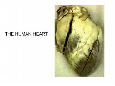
THE HUMAN HEART
Description:
THE HUMAN HEART OVERVIEW OF THE CARDIOVASCULAR SYSTEM Pulmonary circuit Systemic circuit Arteries (Including the coronary arteries) Veins (Including the coronary ... – PowerPoint PPT presentation
Number of Views:148
Avg rating:3.0/5.0
Title: THE HUMAN HEART
1
THE HUMAN HEART
2
OVERVIEW OF THE CARDIOVASCULAR SYSTEM
- Pulmonary circuit
- Systemic circuit
- Arteries (Including the coronary arteries)
- Veins (Including the coronary veins)
- Capillaries (Arterioles Venules)
- Four chambers of the heart
Pulmonary circuit
3
(No Transcript)
4
HEART POSITIONING
- Located near the anterior chest wall
- Posterior to the sternum
- Lies slightly to the left of the midline
- Sits at an angle
- Rotated toward the left side
5
Anatomical position of the heart from Grays
Anatomy
6
Pericardial Cavity
- Anterior cavity of the mediastinum
- Separates the two pleural cavities
- Contains the thymus, esophagus the trachea
7
Pericardium
- Pericardial cavity is lined by the pericardium.
- Visceral pericardium (epicardium) covers the
hearts outer surface. - Parietal pericardium lines the inner surface of
the pericardial sac that surrounds the heart. - Pericardial fluid acts as a lubricant reducing
friction.
8
The clear tissue being Lifted up by the scalpel
Is the pericardium
9
Pericarditis
- Various pathogens may infect the pericardium.
- The inflamed pericardial surfaces rub against one
another. - Makes a distinct scratching sound.
- Cardiac tamponade could occur due to the
increased pericardial fluid in the pericardial
cavity. This condition restricts the movement of
the heart.
10
Black looking structure is the heart bulging from
the pericardial sac. Not only pathogens can
cause a cardiac tamponade, but blunt force trauma
can also cause it.
11
Superficial Heart Anatomy
- When not filled with blood, the outer portion of
each atrium deflates and becomes a lumpy,
wrinkled flap. - This extension is called the auricle (looks like
an external ear). - The coronary sulcus, marks the boundary between
the atria and ventricles.
12
Even though this is the posterior view of the
heart, the coronary sulcus goes around the
entire heart to separate the upper and lower
chambers.
13
The anterior and posterior interventricular sulci
are shallower depressions that mark the boundary
line between the left and right
ventricles. These areas usually contain a large
amount of fat. The sulci contain the arteries and
veins that feed the heart. The heart has an
attached base and a free apex. The inferior tip
is called the apex. In a typical adult the heart
measures approximately 12.5 cm from the base to
the tip. The apex reaches to the fifth
intercostal space, 7.5 cm to the left of the
midline.
14
Interventricular septum
15
Superior Vena Cava. Brings blood from the head,
neck and shoulders to the right atrium
Interatrial septum
Inferior Vena Cava Brings blood back to the right
atrium from the rest of the body
16
Interatrial Septum. Lateral view
17
The pectinate muscles are prominent muscular
ridges found in the atrial walls
18
Blood Flow
- Right atrium into the right ventricle via the
right atrioventricular (AV) valve also called the
tricuspid valve. - The opening is bounded by three fibrous tissue
cusps, therefore called the tricuspid valve. - This tissue is braced by the tendinous chordae
tendineae connected to papillary muscles
19
1
2
3
Chordae tendineae
Papillary muscle
20
Tricuspid Valve
21
Right Heart
- Blood leaving the right ventricle enters the
pulmonary trunk passing through the pulmonary
semilunar valve. - The pulmonary trunk divides into the left and
right pulmonary arteries. - These arteries are the only arteries in the body
that carries oxygen poor blood. - This blood is then carried to the lungs for
re-oxygenation and removal of carbon dioxide.
22
Remember you are looking at the right side of the
heart.
23
5 are the pulmonary semilunar valve. 11 is the
pulmonary trunk.
24
The left and right pulmonary returns the blood to
the left atrium. These are the only veins in the
body that carries oxygen rich blood. Blood will
then pass from the left atrioventricular valve
(AV) or also called the bicuspid or mitral
valve. From the left ventricle blood will then
pass through the aortic semilunar valve into the
ascending aorta into the systemic circulatory
system
25
Pulmonary Veins
26
(No Transcript)
27
(No Transcript)
28
8 is the aortic semilunar valve
29
Ventricular Differences
- The anatomical differences between the right and
left ventricles are as follows The right
ventricle is relatively thin. The left ventricle
has a massive muscular wall.
30
Left Ventricle Heart Wall
Right Ventricle Heart Wall
31
Atrioventricular Valves
- Prevent backflow of blood from the ventricles
back into the atria. - Chordae tendineae and papillary muscles play an
important role in this process. - Ventricular diastole the ventricles relax and the
ventricles refill. - The chorae tendineae are loose and offer no
resistance to the flow of blood.
32
During ventricular systole the ventricles begin
to contract blood moving back towards the atria
swings the cusps together closing the
valves. The chordae tendineae and papillary
muscles stops the cusps from swinging into the
atria. If those two structures are cut or
damaged the valves act as swinging doors, and
there is backflow, or regurgitation. Mitral
valve damage can especially occur in women after
pregnancy.
33
The Heart Wall
- Bulk of the heart consists of the muscular
myocardium and endocardium, that covers the inner
surface of the heart. - The epicardium is the visceral pericardium that
covers the outer surface of the heart.
34
(No Transcript)
35
- Cardiac muscle cells are interconnected by
intercalated discs which convey the force of
contraction from cell to cell and conduct action
potentials.
36
(No Transcript)
37
Heart Blood Supply
- Coronary circulation demands high oxygen and
nutrients for the cardiac muscle cells. - Coronary arteries originate at the base of the
ascending aorta. - Interconnections between arteries called
anastomoses ensure a constant blood supply.
38
Anastomoses
39
(No Transcript)
40
Great, Posterior, small, Anterior, Middle
Cardiac Veins carry blood from The coronary
capillaries To the coronary sinus. Left coronary
artery supplies The left ventricle.
Circulflex Curves left meeting with The right
coronary artery. Left anterior decending Supplies
the posterior Decending artery (interventricular)
.
Recommended
CrystalGraphics Presentations


