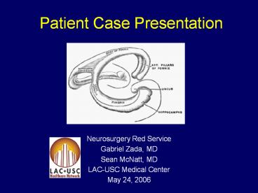
Patient Case Presentation - PowerPoint PPT Presentation
1 / 109
Title:
Patient Case Presentation
Description:
Patient Case Presentation Neurosurgery Red Service Gabriel Zada, MD Sean McNatt, MD LAC-USC Medical Center May 24, 2006 Patient J.P. History of Present Illness: 22 ... – PowerPoint PPT presentation
Number of Views:367
Avg rating:3.0/5.0
Title: Patient Case Presentation
1
Patient Case Presentation
- Neurosurgery Red Service
- Gabriel Zada, MD
- Sean McNatt, MD
- LAC-USC Medical Center
- May 24, 2006
2
Patient J.P.
- History of Present Illness
- 22 year old caucasian female
- Long history of headaches
- Presented with 2 days of
- Sinus headache progressing to
- Bifrontal headache
- Somnolence
- Altered mental status
- Nausea/vomiting
- No fevers, chills
- No history of trauma
3
Patient J.P.
- Past Medical History
- Headaches x 2 years
- Otherwise unremarkable past medical history
- Medications
- None
- Allergies
- None Known
- Social History
- Mother of a 2 year old child
- No tobacco, drug, or alcohol use
4
Patient J.P.
- Physical Exam
- Mental Status
- Patient somnolent, partially arousable
- Oriented inconsistently to name only
- Responds inappropriately with one word responses
- Cranial Nerve Exam
- Right partial ptosis
- Papilledema
- Right pupil 5?4 mm, sluggish
- Left pupil 5?3 mm, brisk
- Extraocular movements intact
- Cranial Nerves otherwise intact
5
Patient J.P.
- Motor exam
- Normal tone
- Follows simple commands intermittently
- Squeezes hands, wiggles toes
- Diffusely weak in all extremities
- Sensory Exam
- Sensation intact to light touch in all
extremities - Reflexes
- Reflexes 2, symmetrical
- No Hoffmans sign
- Toes downgoing
- Cerebellar/Gait exam
- Mild dysmetria bilaterally on finger-nose test
- Gait Deferred
6
CT Scan
7
(No Transcript)
8
(No Transcript)
9
(No Transcript)
10
(No Transcript)
11
(No Transcript)
12
(No Transcript)
13
(No Transcript)
14
(No Transcript)
15
(No Transcript)
16
Initial Management
- Patient transferred to LAC Medical Center
- Right ventriculostomy placed
- CSF sent for cytology ? atypical cells
- Patients exam significantly improved
- Awake, alert
- Oriented to name only (San Dimas , 1993)
- Significant short-term memory deficits
- Partial right IIIrd nerve palsy improved
- No pronator drift, power 5/5 throughout
17
CT Scan
18
(No Transcript)
19
(No Transcript)
20
(No Transcript)
21
(No Transcript)
22
(No Transcript)
23
(No Transcript)
24
(No Transcript)
25
(No Transcript)
26
(No Transcript)
27
MRI Brain
28
(No Transcript)
29
(No Transcript)
30
(No Transcript)
31
(No Transcript)
32
(No Transcript)
33
(No Transcript)
34
(No Transcript)
35
(No Transcript)
36
(No Transcript)
37
(No Transcript)
38
(No Transcript)
39
(No Transcript)
40
(No Transcript)
41
(No Transcript)
42
(No Transcript)
43
(No Transcript)
44
(No Transcript)
45
(No Transcript)
46
(No Transcript)
47
(No Transcript)
48
(No Transcript)
49
(No Transcript)
50
(No Transcript)
51
(No Transcript)
52
(No Transcript)
53
(No Transcript)
54
(No Transcript)
55
(No Transcript)
56
(No Transcript)
57
(No Transcript)
58
(No Transcript)
59
(No Transcript)
60
(No Transcript)
61
Surgery
- Right interhemispheric transcallosal approach to
third ventricle - Patients right side down
- Frozen pathology ? malignant glial tumor with
high cellularity - Gross total resection
62
(No Transcript)
63
(No Transcript)
64
(No Transcript)
65
(No Transcript)
66
Transcallosal Approach to the Third Ventricle
- Position with head in lateral position to allow
gravity to facilitate in retraction - Bone flap 2/3 anterior to coronal suture and 1/3
posterior to coronal suture - May modify accordingly for anterior versus
posterior third ventricular lesions - Callosal incision between 2 ACAs
- Must account for shift involved with lateral
positioning - Callosotomy approximately 2-3 cm in length
- Some authors advocate transverse callosotomy
67
Video
68
Postoperative MRI Brain
69
(No Transcript)
70
(No Transcript)
71
(No Transcript)
72
(No Transcript)
73
(No Transcript)
74
(No Transcript)
75
(No Transcript)
76
(No Transcript)
77
(No Transcript)
78
(No Transcript)
79
(No Transcript)
80
(No Transcript)
81
(No Transcript)
82
(No Transcript)
83
(No Transcript)
84
(No Transcript)
85
(No Transcript)
86
(No Transcript)
87
(No Transcript)
88
(No Transcript)
89
(No Transcript)
90
(No Transcript)
91
(No Transcript)
92
Postoperative Course
- Patient with unchanged neurological status
following procedure - Ventriculostomy left in place yet unable to wean
off - Left VP shunt placed on postoperative day 7
- Short term memory slightly improved over course
of week - Patient transferred to step-down
93
Pathology
94
(No Transcript)
95
(No Transcript)
96
(No Transcript)
97
(No Transcript)
98
DiagnosisIntraventricular Anaplastic
Oligodendroglioma (WHO Grade III)
99
Oligodendroglioma Background
- Two recognized grades
- WHO grade II oligodendroglioma
- WHO grade III anaplastic oligodendroglioma
- 4 of all primary brain tumors
- Mean age approximately 43 years
- 6 during infancy and childhood
- No known patterns of inheritance
- Most commonly occur in white matter of frontal
and temporal lobes - Intraventricular oligodendroglioma
- Approximately 20 case reports in the literature
100
Anaplastic Oligodendroglioma Epidemiology
- Account for 3 of all adult supratentorial
primary malignant gliomas - Account for 20-54 of all oligodendrogliomas
- Most common in adults (mean age 49 years)
- Older than patients with grade II
oligodendrogliomas - Male to female ratio 1.5 1
- Preference for frontal lobe (60) followed by
temporal lobe (33)
101
Anaplastic Oligodendroglioma Histopathology
- Share with oligodendroglioma
- Honeycomb appearance with clear cytoplasm
- Fried egg yolk appearance
- Frequent calcification
- Occasional gemistocytes
- Often GFAP and S-100 positive
- Perinuclear halos
- Diffuse features of malignancy
- Increased cellularity
- Cellular atypia
- High mitotic index
- Necrosis and microvascular proliferation may be
present - Occasional multinucleated giant cells of Zulch
102
Anaplastic Oligodendroglioma Differential
Diagnosis
- All with neoplastic cells with round nucleus and
clear cytoplasm (oligodendroglioma-like cells or
OLCs) - Clear cell ependymoma
- ependymal features (ie rosettes) help
differentiate - Central neurocytoma
- Synaptophysin positive, more commonly originates
in ventricles - Clear cell meningioma
- PAS positive, EMA immunoreactivity
- Metastatic renal cell tumor
103
Oligodendroglioma Molecular Genetics
- Chromosome 19
- Loss of heterozygosity (LOH) on long arm of
chromosome 19 (19q) - 50-80 of cases
- Chromosome 1
- LOH on short arm (1p) in 67 of cases
- Almost always coexists with LOH at 19q
- Polysomia, deletions on other chromosomes (ie
9,10) - Progression to malignancy correlates with EGFR,
PDGF overexpression - Fluorescence In Situ Hybridization (FISH) used to
detect - Lack of correlation between histology and
molecular markers
104
Oligodendroglioma Molecular genetics
- Several molecular subtypes
- 1) Combined/isolated loss of 1p/19q
- More likely frontal, parietal
- Diffuse enhancement
- Close to 100 response rate
- Survival greater than 10 years
- 2) 1p loss without 19q loss
- Close to 100 response rate
- Survival approximately 6 years
- 3) No deletion of 1p/19q with TP53 mutation
- More likely temporal, insular
- Ringe enhancement more likely
- 33 response rate
- Survival approximately 6 years
- 4) No deletion of 1p/19q, no TP53 mutation, yet
other mutations - 18 response rate
- Survival generally less than 18 months
105
Anaplastic Oligodendroglioma Multimodal treatment
- Surgery is still primary treatment
- Gross total resection whenever possible
- Mixed data regarding adjuvant radiotherapy
- Postoperative radiation therapy has been shown to
extend survival, yet carries associated morbidity - Delayed XRT as effective as immediate postop XRT
in one study - Another study showed no benefit to radiotherapy
- XRT/chemo current standard in recurrent,
high-grade oligodendroglioma - Salvage therapy frequently chemotherapy with stem
cell rescue
106
Anaplastic Oligodendroglioma Response to
chemotherapy
- Tumors with combined 1p and 19q deletions are
often responsive to chemotherapy - Procarbazine, CCNU, Vincristine (PCV)
- Many side effects including myelosuppression in
46 - More recently, temozolamide (in trials)
- Half of such tumors show complete radiological
responses to chemotherapy - Mean survival time 10 years with these deletions
compared to patients without these deletions
(mean 2 years)
107
Anaplastic Oligodendroglioma Prognosis
- Median survival time of 4 years
- Five year survival 41
- Ten year survival 20
- Local tumor recurrence occurs frequently
- Leptomeningeal spreading (oligodendrogliomatosis
) has also been described - Metastatic disease uncommon, yet incidence may be
increasing - Most common sites bone, lymph nodes, scalp
- Good prognostic factors
- Younger patient age
- Female sex
- Seizure as presenting symptom
108
References
- 1. Merrell R et al. 1p/19q chromosome deletions
in metastatic oligodendroglioma. J Neurooncology.
2006 - 2. Waldron JS, Tihan T. Epidemiology and
pathology of intraventricular tumors.
Neurosurgery Clinical of N America. 14 (2003)
469-482 - 3. Dumont AS et al. Intraventricular gliomas.
Neurosurgery Clinical of N America. 14 (2003)
571-591 - 4. Reifenberger G. Anaplastic oligodendroglioma.
In Tumours of the Nervous System. (Kleihues P,
Cavanee WK, eds.) IARC Press, 2000. - 5. Kasowski HJ et al. Transcallosal
Transchoroidal Approach to Tumors of the Third
Ventricle. Neurosurgery 57, Suppl 3. 361-366,
2005 - 6. Engelhard HH. Current diagnosis and treatment
of Oligodendroglioma. Neurosurgical Focus. 12(2),
2002.
109
Thank You


