
Hybrid Convolution Kernel: Optimized CT of the Spine - PowerPoint PPT Presentation
1 / 1
Title:
Hybrid Convolution Kernel: Optimized CT of the Spine
Description:
Hybrid Convolution Kernel: Optimized CT of the Spine Kenneth L Weiss MD*, Aaron L Greeley DO, Rebecca S Cornelius MD, Dongmei Sun PhD Department of Radiology ... – PowerPoint PPT presentation
Number of Views:47
Avg rating:3.0/5.0
Title: Hybrid Convolution Kernel: Optimized CT of the Spine
1
Hybrid Convolution Kernel Optimized CT of the
Spine
Kenneth L Weiss MD, Aaron L Greeley DO, Rebecca
S Cornelius MD, Dongmei Sun PhD
Department of Radiology, University of
Cincinnati, Cincinnati, Ohio
Introduction
Results
Selection of a CT convolution kernel determines
the tradeoff between image sharpness and pixel
noise.(1) High pass filter algorithms utilized in
commercially available sharp convolution
kernels preserve higher spatial frequencies at
the expense of greater noise and typically work
best for tissues with inherently high CT
contrast. Conversely, low pass algorithms
utilized in smooth convolution kernels reduce
the higher frequency contribution decreasing
noise and spatial resolution and work best for
tissues with inherently lower contrast such as
the brain or liver.(2, 3) Consequently, as most
clinical exams include tissues with both high and
low inherent contrast, it is often desirable to
create at least two separate data sets utilizing
different convolution kernels. Unfortunately,
this increases the number of images needed to be
generated, transmitted, stored and reviewed by a
corresponding factor of two or more. To address
this problem, Schaller et al. described a spatial
domain filtering algorithm for fast modification
of the image sharpness-pixel noise tradeoff.
While providing the ad hoc ability to reduce the
noise and spatial resolution of images generated
with a high pass convolution kernel, tradeoffs
exist and the resultant images only approximate
those prospectively created with routine low pass
convolution kernels.(1) Rather than develop a
distinct algorithm to approximate routine
clinical convolution kernels, we chose to combine
well established kernels in such a fashion as to
directly duplicate within a single hybrid image
the established tissue contrast that had been
individually optimized for soft tissues or bone.
In so doing, we hoped to halve the number of
images to be archived and reviewed without
compromising or altering clinically established
CT tissue contrast thereby obviating a learning
curve and facilitating comparison with
conventional single kernel images.
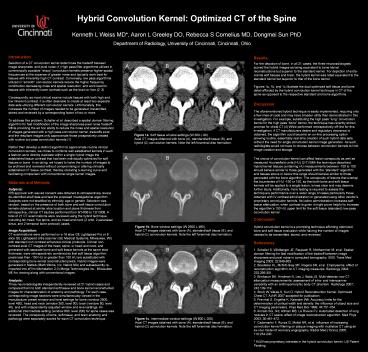
For the depiction of bone, in all 21 cases, the
three neuroradiologists scored the hybrid images
as being equivalent to bone kernel
reconstructions but superior to the standard
kernel. For depiction of extra-cranial soft
tissues and brain, the hybrid kernel was rated
equivalent to the standard kernel but superior to
that of the bone kernel. Figures 1a, 1b, and 1c
illustrate the dual optimized soft tissue and
bone detail afforded by the hybrid convolution
kernel technique in CT of the spine as opposed to
the respective standard and bone algorithms.
Discussion
The aforementioned hybrid technique is easily
implemented, requiring only a few lines of code
and may have broader utility than demonstrated in
this investigation. For example, substituting the
high pass lung convolution kernel for the high
pass bone kernel, the technique has recently
shown promise for chest CT.(4) While performed
retrospectively off-line for this investigation,
if CT manufacturers desire and regulatory
clearance is obtained, the algorithm could become
an on-line processing option allowing routine,
essentially real-time creation of such hybrid
data sets without the need for single convolution
kernel image generation. As such, radiologists
would not have to choose between convolution
kernels to limit image creation and storage.
The choice of convolution kernel can affect
lesion conspicuity as well as measured
Hounsefield units (HU).(2-7) With the technique
described, hybrid kernel tissues containing HU
measurements between -150 to 150 should behave
similar to those generated with the standard
algorithm and tissues above or below this range
should behave similar to those generated with the
bone algorithm. The conspicuity of lesions that
overlap the boundaries of HU -150 or 150, so that
both bone and soft tissue kernels will be applied
to a single lesion, is less clear and may deserve
further study. Additionally, more testing is
required to assess the techniques performance
over a wider range of cases particularly those
obtained with IV contrast administration or
generated using other vendors proprietary
convolution kernels. As iodine administration
increases soft tissue attenuation, when contrast
is given it might prove helpful to increase the
algorithms 150 HU upper limit for the soft
tissue (standard) low-pass convolution kernel.
Materials and Methods
Subjects IRB approval with waived consent was
obtained to retrospectively review de-identified
shelf data and test the proposed investigational
algorithm. Subjects were not stratified by
ethnicity, age or gender. Selection was random,
based on the presence of both bone and soft
tissue convolution kernels obtained at similar
slice location and plane thickness from
retrospective, clinical CT studies performed from
9/14/06 to 12/13/06. A total of 21 CT
examinations were reviewed using the hybrid
technique, including ten head, five spine, and
six head and neck (2 orbit, 2 paranasal sinus,
and 2 temporal bone protocol) cases.
Conclusion
Hybrid convolution kernel is a promising
technique affording optimized bone and soft
tissue evaluation while halving the number of
images needed to be transmitted, stored, and
reviewed.
Image Acquisition CT examinations were performed
on a 16 slice GE Lightspeed Pro or 8 slice GE
Lightspeed Ultra scanner (GE Medical Systems,
Milwaukee, WI) with standard non contrast
enhanced clinical protocols. Clinical
non-contrast axial CT images of the head, spine,
or head and neck and generated with separate
bone and soft tissue kernels at the same slice
thickness, were retrospectively combined so that
soft tissue algorithm pixels less than -150 HU or
greater than 150 HU are substituted with
corresponding bone kernel reconstructed pixels.
Hybrid images were generated in Matlab (Math
Works, Inc, Natick MA) and subsequently
re-imported into eFilm Workstation 2.0 (Merge
Technologies Inc., Milwaukee WI) for viewing
along with conventional images.
References
1. Schaller S, Wildberger JE, Raupach R,
Niethammer M, et al. Spatial domain filtering for
fast modification of the tradeoff between image
sharpness and pixel noise in computed tomography.
IEEE Trans Med Imaging 2003 22846-853. 2.
Boedeker KL, McNitt-Gray MF, Rogers SR, et al.
Emphysema effect of reconstruction algorithm on
CT imaging measures. Radiology 2004
232295-301. 3. Birnbaum BA, Hindman N, Lee J,
Babb JS. Multi-detector row CT attenuation
measurements assessment of intra- and
interscanner variability with an anthropomorphic
body CT phantom. Radiology 2007 242109-119. 4.
Strub W, Weiss K, Sun D. Hybrid Reconstruction
Kernel Optimized Chest CT. AJNR 2007accepted
for publication. 5. Prevrhal S, Engelke K,
Kalender WA. Accuracy limits for the
determination of cortical width and density the
influence of object size and CT imaging
parameters. Phys Med Biol 1999 44751-764. 6.
Armato SG, 3rd, Altman MB, La Riviere PJ.
Automated detection of lung nodules in CT scans
effect of image reconstruction algorithm. Med
Phys 2003 30461-472. 7. Cademartiri F, Runza G,
Mollet NR, et al. Influence of increasing
convolution kernel filtering on plaque imaging
with multislice CT using an ex-vivo model of
coronary angiography. Radiol Med (Torino) 2005
110234-240 KLW has proprietary interests in
the hybrid convolution kernel, US Patent Pending
Analysis Three neuroradiologists independently
reviewed all 21 hybrid cases and compared them to
both standard soft tissue and bone kernel
reconstructed images for characterization of
anatomy and pathology. For each case,
corresponding image sections were simultaneously
viewed in the manufacturer preset window and
level settings for bone (window 2500, level 480),
head and neck (window 350, level 90), brain
(window 80, level 40), and with independently
adjusted window and level settings. An additional
intermediate setting (window 800, level 200) for
spine cases was reviewed. The conspicuity of
bone, soft tissue, and brain anatomy and
pathology were separately scored for each CT
convolution technique.