
Agents of Bioterrorism - PowerPoint PPT Presentation
1 / 53
Title:
Agents of Bioterrorism
Description:
Personnel specifically trained in handling pathogenic agents ... 1968 Carmichael - Beagles, B. canis. 52. BRUCELLOSIS: TRANSMISSION. Unpasteurized dairy products ... – PowerPoint PPT presentation
Number of Views:265
Avg rating:3.0/5.0
Title: Agents of Bioterrorism
1
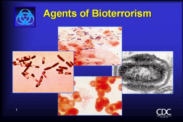
Agents of Bioterrorism
2
Subject Matter Experts at CDC
- Bacillus anthracis - Robbin Weyant
- Brucella spp - Robbin Weyant
- Botulinum toxin - Susan Maslanka
- Francisella tularensis - May Chu
- Hemorrhagic fevers - Tom Ksiazek
- Smallpox - Joe Esposito
- Yersinia pestis - May Chu
3
Level A LaboratoryDefinition
- BSL-2 Laboratory with a certified Class II
biological safety cabinet - BSL-1 microbiology practices plus
- Directed by competent scientists
- Personnel specifically trained in handling
pathogenic agents - Biological safety cabinet, Class II
- Access limited by lab director
4
Level A LaboratoryDefinition, cont
- BSL-2 Laboratory with a certified Class II
biological safety cabinet - Physical containment practices to minimize
infectious aerosols - Sharpsprecautions
- PPE (lab coat, gloves, face shield)
- Biohazard warning signs
- Biosafety manual defining waste/ SH/
decontamination/surveillance policy/ CC
5
Role of the Level A Laboratory
- Rule out critical biological agents
- Refer to higher level laboratory
6
Bioterrorism AgentsLaboratory Risk
- Agent BSL Laboratory Risk
- B. anthracis 2 low
- Y. pestis 2 medium
- F. tularensis 2/3 high
- Brucella spp. 2/3 high
- Botulinum toxin 2 medium
- Smallpox 4 high
- Viral Hemorrhagic fever 4 high
7
Francisella tularensis
- Tularemia
8
Francisella tularensisA Rose by Any Other Name
- Plague-like disease in rodents (California)
- Deer-fly fever (Utah)
- Glandular tick fever (Idaho and Montana)
- Market mens disease (Washington, DC)
- Rabbit fever (Central States)
- OHaras disease (Japan)
- Water-rat trappers disease (Russia)
9
Reported Cases of Tularemia - 1990-1998
10
Level A ProceduresFrancisella tularensis
- This is a dangerous, highly virulent organism and
it should not be manipulated at the bench.
Laboratory-acquired infections can occur easily. - Gram stain
- Growth characteristics in broth
- Growth characteristics in agar
11
Francisella tularensis
- Gram stain
- Poorly staining, tiny gram-negative
- coccobacilli
12
Francisella tularensisGrowth Characteristics
- Fastidious, requires cysteine for robust growth
Cysteine Heart Agar (CHA) is ideal - Enriched chocolate agar 9 sheep blood
cysteine - Not part of Level A routine procedures
- BCYE (for Legionella) also works
13
Francisella tularensisGrowth Characteristics
- Will grow initially on sheep and chocolate blood
agar and - Thayer-Martin agar, but poorly or not at all
on passage - Grows slowly at 35oC, poorly at 28oC
14
Francisella tularensisGrowth Characteristicscont
inued
- 24 hours on SBA, CA, TM, CHA
- gray-white, translucent colonies
- usually too small to be seen individually
15
Francisella tularensisGrowth Characteristicscont
inued
- 48 hours on SBA, CA, TM, CHA
- SBA - lt1 mm, gray-white, opaque, no hemolysis
- TMA, CA - 1-2 mm,gray-white, flat, entire,
smooth, shiny - CHA - 2-4 mm, greenish-white, dense, shiny,
opalescent sheen
16
Gram Negative Coccobacilli
- Most likely
- Acinetobacter (ox.neg)
- Actinobacillus (sticky)
- H. aphrophilus
- Bordetella, Grp. IV (inert, urea pos)
- Pasturella (nonsticky, Mac pos)
- Least likely
- DF-3
- Brucella (Urea pos in seconds - minutes)
- Francisella (Urea neg)
17
Francisella tularensisRapid Method Results
- Not on the data base of MicroScan or Vitek or API
- Should not be worked with in the Level A lab
18
Tularemia
- Contagious --- no
- Infective dose --- 10-50 organisms
- Incubation period --- 1-21 days (average3-5
days) - Duration of illness --- 2 weeks
- Mortality --- treated low untreated
moderate - Persistence of organism ---months in moist soil
- Vaccine efficacy --- good, 80
19
Francisella tularensisTechnical Hints
If you see
- Tiny, gram-negative coccobacilli from blood,
lymph node aspirate, or respiratory specimens - Blood isolates that will grow slowly on chocolate
agar but poorly or not at all on blood agar in 24
hours - Faint growth in thio requires cysteine in other
broth
Refer
20
Yersinia pestis
- Plague
21
Plague Epidemiology
- U.S. averages 13 cases/yr (10 in 1998)
- 30 of cases are in Native Americans in the
Southwest. 15 case fatality rate
22
Plague Epidemiologycontinued
- Most cases occur in summer and near the patients
residence - bubonic (infected lymph nodes)
- septicemic (blood-borne organisms)
- pneumonic (transmissible by aerosol deadliest)
23
Yersinia pestisSpecimen Selection
- Specimen selection is important
- Bubonic - bubo - lymph node aspirate
- Septicemic - blood - organisms may be
intermittent. Take three specimens 10-30 minutes
apart - Pneumonic
- Sputum/throat - use Wayson and DFA stain
- Bronchial washings - Wayson and DFA stain
24
Yersinia pestisspecimen inoculation
- Inoculate routine plating media and make thin
smear for DFA - Use Wayson only if DFA is unavailable
25
Level A ProceduresYersinia pestis
- Gram stain
- Wayson stain
- Growth characteristics on agar
- Growth characteristics in broth
26
Yersinia pestisGram stain
- Small, gram-negative bipolar-stained coccobacilli
Must confirm by DFA and mouse inoculation
27
Yersinia pestisWayson Stain
- Used for rapid assessment
- for specimens when DFA is not available
- when it is a part of the identification process
- Best with tissue, sputum, blood
- Stains of pure culture isolates tend to lose
bipolarity
28
Yersinia pestisWayson Stain
- Pink-blue cells with a closed safety pin look
Wayson stain alone is not diagnostic
29
Yersinia pestisGrowth in Broth
- Brain Heart Infusion Broth (two tubes)
- Incubate at 28oC (best) and 37oC for
- 24-48h
- Do not shake tubes
- Observe suspended flocculent clumps like
stalactites on side and bottom of tube. Broth
remains clear
30
Yersinia pestis in Broth
Y. pestis
Y. pseudotuberculosis
31
Yersinia pestis Growth on Agar
- Sheep blood agar - 28oC (faster)
- and 37oC (for DFA tests)
- Looks like other enterics
32
Yersinia pestisRapid Method Results
- On the data base of MicroScan, Vitek, and API 20E
- True accuracy not yet determined
33
Yersinia pestisTechnical Hints
- Small gram-negative, poorly staining rods from
blood, lymph node aspirate, or respiratory
specimens - Safety pin appearance in Gram, Wright, Giemsa, or
Wayson stain - More than one patient in a short, specified
period with fever, lymphadenopathy
Refer
34
Variola virus
- Smallpox
35
VariolaSmallpox virus
- Large DNA virus
- Dumbbell-shaped core
- Complex membranes
Refer
36
VariolaSmallpox virus
- The family Poxviridae consists of eight genera
and a few unclassified species - Two species are human viruses
- Variola virus (genus Orthopoxvirus)
- Molluscum contagiosum virus (genus
Molluscipoxvirus) - Orthopoxvirus includes vaccinia (a lab virus),
monkeypox, cowpox, and buffalopox
37
Smallpox virus
- Stored stocks to be retained until 2002 by U.S.
and Russia - Undeclared virus could be anywhere
- No cases in over 20 years
- Controversial decision
- Immunity lost in U.S. population
- Highly vulnerable to infection
38
Level A ProceduresSmallpox virus
- Rule out chickenpox (PCR)!
- Specimen of choice is lesion material from
pustules. - Collect vesicular fluid from each single lesion
- Place droplet fluid as a drop on a clean slide -
Do not smear - Store each slide in separate slide holder
- Capillary tubes or dry swabs are alternatives
39
Level A ProceduresSmallpox virus
- Autopsy specimens must be frozen.
Formalin fixation is OK for histopathologic study - Contact CDC for approval to ship
- Send slide in a non-breakable holder. Do not use
a transport fluid. - Store at 4oC briefly or at -20oC to -70oC
- Decontaminate with 0.5 hypochlorite
40
Hemorrhagic Fever Viruses
Marburg
Ebola
41
Hemorrhagic Fever Viruses
- Arenaviruses
- Argentine HF
- Bolivian HF
- Sabia Associated HF
- Lassa fever
- Lymphocytic choriomeningitis
- Venezuelan HF
- Bunyaviruses
- Crimean-Congo HF
- Rift Valley Fever
- Hantavirus Pulmonary Syndrome HF with 0Renal
Syndrome
42
Hemorrhagic Fever Viruses (continued)
- Filoviruses
- Ebola HF
- Marburg HF
- Flaviviruses
- Tick-borne Encephalitis
- Kyasanur Forest Disease Omsk HF
43
Viral Hemorrhagic Fevers
- Contagious --- Moderate
- Infective dose --- 1-10 particles
- Incubation period --- 4-21 days
- Duration of illness --- 7-16 days
- Mortality ---variable
- Persistence of organism --- unstable
- Non-endemic in U.S.
- Vaccine efficacy --- no vaccine
44
VHF Specimens
- Diagnosis is clinical, not laboratory
Refer
45
Handling VHF Specimens
- No specimen accepted without prior consultation -
404-639-1115 - Serology - 10-12 ml 5 ml minimum
- serum drawn at admission (acute, convalescent 21
days later post-mortem heart blood - ship serum cold or on dry ice in a plastic tube
46
Handling VHF Specimens-Immunohistochemistry
- Prefer lung, kidney, spleen tissue
- Other lymph node, heart, pancreas,
- pituitary, brain, liver
- Paraffin blocks preferred. Formalin-fixed tissue
acceptable - Ship blocks/tissue at RT - do not freeze.
Autopsy/surgical report required.
47
Handling VHF Specimens-PCR/Virus isolation
- Ante-mortem - biopsy of lung or bone marrow
aspirate or clot - Post-mortem - spleen, lung, kidney, liver, nodes,
heart, pancreas, pituitary, brain, liver - Must be at least 1 cm3
- Buffy coat, clot, tissue - dry ice
48
Brucella spp.
- Brucellosis
49
BRUCELLOSIS
- A zoonotic disease caused by any of 4 Brucella
sp. abortus, melitensis, suis, and canis - A systemic infection characterized by an undulant
fever pattern - But relatively rare in the U.S. with
approximately 100 cases/yr
50
(No Transcript)
51
BRUCELLOSIS HISTORY
- 1887 Bruce - Malta fever, M. melitensis
- 1897 Bang - cattle abortion, B. abortus
- 1914 Traum - sow, B. suis
- 1920 Evans, Meyer, Shaw - Brucella
- 1954 B. suis, first weaponized U.S. agent
- 1968 Carmichael - Beagles, B. canis
52
BRUCELLOSISTRANSMISSION
- Unpasteurized dairy products
- The most common mode of transmission
- Direct skin contact
- Occupational hazard for farmers, butchers,
veterinarians, and laboratory personnel - Aerosols
- Highly infectious
53
BRUCELLOSIS
- Infective dose 10 -100 organisms
- Incubation period 5 days - gt 6 months
- Duration of illness weeks to months
- Fever, profuse sweating, malaise, headache and
muscle/back pain. - Person to person transmission no
- Mortality lt5
- Persistence of organism very stable
Recommended
CrystalGraphics Presentations


