
Insect Reproduction PowerPoint PPT Presentation
Title: Insect Reproduction
1
Insect Reproduction Development
2
The reproductive organs of insects are similar in
structure and function to those of vertebrates
a male's testes produce sperm and a female's
ovaries produce eggs (ova). Both types of
gametes are haploid and unicellular, but eggs are
usually much larger in volume than sperm. Most
insect species reproduce sexually -- one egg from
a female and one sperm from a male fuse (syngamy)
to produce a diploid zygote. But there are also
many species that reproduce by parthenogenesis,
asexual reproduction in which there is growth and
development of an unfertilized egg. Some
species alternate between sexual and asexual
reproduction (not all generations produce males),
others are exclusively parthenogenetic (no males
ever occur).
3
Male Reproductive System
The male's reproductive system contains a pair of
testes, usually located near the back of the
abdomen. Each testis is subdivided into
functional units (called follicles) where sperm
are actually produced. A typical testis may
contain hundreds of follicles, generally aligned
parallel to one another. Near the distal end of
each follicle, there are a group of germ cells
(spermatogonia) that divide by mitosis and
increase in size to form spermatocytes. These
spermatocytes migrate toward the basal end of the
follicle, pushed along by continued cell division
of the spermatogonia. Each spermatocyte undergoes
meiosis this yields four haploid spermatids
which develop into mature spermatozoa as they
progress further along through the follicle.
4
Mature sperm pass out of the testes through short
ducts (vasa efferentia) and collect in storage
chambers (seminal vesicles) that are usually
little more than enlarged sections of the vasa.
Similar ducts (vasa deferentia) lead away from
the seminal vesicles, join one another near the
midline of the body, and form a single
ejaculatory duct that leads out of the body
through the male's copulatory organ (called an
aedeagus). One or more pairs of accessory glands
are usually associated with the male's
reproductive system. These are secretory organs
that connect to the reproductive system by means
of short ducts -- some may attach near the testes
or seminal vesicles, others may be associated
with the ejaculatory duct. The glands have two
major functions 1. Manufacture of seminal
fluid, a liquid medium that sustains and
nourishes mature sperm while they are in the
male's genital system. 2. Production of
spermatophores, pouch-like structures (mostly
protein) that encase the sperm and protect them
as they are delivered to the female's body during
copulation.
5
Female Reproductive System
The female's reproductive system contains a pair
of ovaries. When the insect is actively
reproducing, these organs swell with developing
eggs and may nearly fill the abdomen. Each
ovary is subdivided into functional units (called
ovarioles) where the eggs are actually produced.
A typical ovary may contain dozens of
ovarioles, generally aligned parallel to one
another. Near the distal end of each ovariole,
there are a group of germ cells (oogonia) that
divide by mitosis and increase in size to form
oocytes. During active oogenesis, new oocytes are
produced on a regular schedule within each
ovariole. These oocytes migrate toward the
basal end of the ovariole, pushed along by
continued cell division of the oogonia. Each
oocyte undergoes meiosis this yields four
cells -- one egg and three polar bodies. The
polar bodies may disintegrate or they may
accompany the egg as nurse cells. .
6
As developing eggs move down the ovariole, they
grow in size by absorbing yolk (supplied by
adjacent nurse cells or accessory cells). Thus,
each ovariole contains a linear series of eggs in
progressive stages of maturation, giving the
appearance of a "chain of beads" where each bead
is larger than the one behind it. By the time
an egg reaches the base (calyx) of the ovariole
it has reached full size -- often growing up to
100,000 times larger than the original oocyte.
Mature eggs leave the ovaries through short
lateral oviducts. Near the midline of the body,
these lateral oviducts join to form a common
oviduct which opens into a genital chamber called
the bursa copulatrix. Female accessory glands
(one or more pairs) supply lubricants for the
reproductive system and secrete a protein-rich
egg shell (chorion) that surrounds the entire
egg. These glands are usually connected by
small ducts to the common oviduct or the bursa
copulatrix.
7
During copulation, the male deposits his
spermatophore in the bursa copulatrix.
Peristaltic contractions force the spermatophore
into the female's spermatheca, a pouch-like
chamber reserved for storage of sperm. A
spermathecal gland produces enzymes (for
digesting the protein coat of the spermatophore)
and nutrients (for sustaining the sperm while
they are in storage). Sperm may live in the
spermatheca for weeks, months, or even years!
During ovulation, each egg passes across the
opening to the spermatheca and stimulates release
of a few sperm onto the egg's surface. These
sperm swim through the micropyle (a special
opening in the egg shell) and get inside the egg.
Fertilization occurs as soon as one sperm's
nucleus fuses with the egg cell's nucleus.
Oviposition (egg laying) usually follows closely
after fertilization. Once these processes are
complete, the egg is ready to begin embryonic
development.
8
Egg Structure
In most insects, life begins as an independent
egg. This type of reproduction is known as
ovipary. Each egg is manufactured within the
female's genital system and is eventually
released from her body through an ovipositor, a
tube-like, saw-like, or blade-like component of
her external genitalia. Production of eggs by
the female's body is called öogenesis and the
egg-laying process is known as oviposition.
Each insect species produces eggs that are
genetically unique and often physically
distinctive as well -- spherical, ovate, conical,
sausage-shaped, barrel-shaped, or torpedo-shaped.
Yet regardless of size or shape, each egg is
composed of only a single living cell -- the
female gamete.
9
An egg's cell membrane is known as the vitelline
membrane . It is a phospholipid bilayer similar
in structure to most other animal membranes. It
surrounds the entire contents of the egg cell,
most of which consists of yolk (food for the
soon-to-develop embryo). The cell's cytoplasm
is usually distributed in a thin band just inside
the vitelline membrane (where it is commonly
called periplasm ) and in diffuse strands that
run throughout the yolk ( cytoplasmic reticulum
). The egg cell's nucleus (haploid) lies within
the yolk, usually close to one end of the egg.
Near the opposite end, the öosome (a region of
higher optical density) may be visible as a dark
region in the more translucent yolk. The egg's
anterior/posterior polarity is determined by the
relative positions of the nucleus and the öosome.
In most insects the egg is covered by a
protective "shell" of protein secreted before
oviposition by accessory glands in the female's
reproductive system. This egg shell, called the
chorion , is often sculptured with microscopic
grooves or ridges that may be visible only under
the high magnification of an electron microscope.
The chorion is perforated by microscopic pores
(called aeropyles ) that allow respiratory
exchange of oxygen and carbon dioxide with
relatively little loss of water. The micropyle
, a special opening near the anterior end of the
chorion, serves as a gateway for entry of sperm
during fertilization.
10
A female receives sperm from her male partner
during the act of mating. She can store that
sperm for long periods of time in a special part
of her reproductive system, the spermatheca. As
a developing egg moves past the opening to the
spermatheca, a few sperm are released onto its
surface. The sperm swim toward the micropyle --
the first one to reach its destination enters and
injects its nucleus into the egg. The sperm
nucleus quickly fuses with the egg nucleus
(syngamy) to form a diploid zygote -- a
one-celled embryo. This event is known as
fertilization. After the egg is fertilized, it
undergoes a period of rapid growth and
development known as embryogenesis, literally the
"embryo's beginning".
11
Embryogenesis
Embryogenesis is a developmental process that
usually begins once the egg has been fertilized.
It involves multiplication of cells (by
mitosis) and their subsequent growth, movement,
and differentiation into all the tissues and
organs of a living insect. The field of insect
embryology has recently yielded stunning insights
into the developmental processes of humans and
other vertebrate organisms. There is remarkable
similarity in genes responsible for organizing
the fundamental body plan in vertebrates and
invertebrates. For example, eyeless, a gene
needed for development of an insect's compound
eyes is also necessary for development of a
mouse's vertebrate eyes! Although much of insect
embryology is still a mystery, there has been
remarkable progress in knowledge over the past
few years thanks to new methods in molecular
biology and genetic engineering. Fruit flies,
silkworms, and hornworms are proving to be a
"rosetta stone" for embryology.
12
An insect's egg is much too large and full of
yolk to simply divide in half like a human egg
during its initial stages of development (imagine
how much time and energy it would take just to
build new cell membranes!). Birds have this
same problem -- think of the yolk in a chicken's
egg. Birds solve the problem by having the
embryo develop within a tiny spot of cytoplasm
(the blastodisc) on the surface of the yolk.
Insects solve the problem by "cloning" the zygote
nucleus (mitosis without cytokinesis) through
12-13 division cycles to yield about 5000
daughter nuclei. This process of nuclear
division is known as superficial cleavage (in
"true" cleavage entire cells divide). As they
form, the cleavage nuclei (often called
"energids") migrate through the yolk toward the
perimeter of the egg. They settle in the band
of periplasm where they engineer the construction
of membranes to form individual cells. The end
result of "cleavage" is the blastoderm -- a
one-cell-thick layer of cells surrounding the
yolk.
13
The first cleavage nuclei to reach the vicinity
of the öosome are "reserved" for future
reproductive purposes -- they do not travel to
the periplasm and do not form any part of the
blastoderm. Instead, they stop dividing and
form germ cells that remain segregated thoughout
much of embryogenesis. These cells will
eventually migrate into the developing gonads
(ovaries or testes) to become primary öocytes or
spermatocytes. Only when the adult insect
finally reaches sexual maturity will these cells
begin dividing (by meiosis) to form gametes of
the next generation (eggs or sperm). Germ cells
never grow or divide during embryogenesis, so DNA
for the next generation is "conserved" from the
very beginning of development. This strategy
has a clear selective advantage it minimizes
the risk that an error in replication (a genetic
defect) will accidently be passed on to the next
generation. Blastoderm cells on one side of the
egg begin to enlarge and multiply. This region,
known as the germ band (or ventral plate), is
where the embryo's body will develop. The rest
of the cells in the blastoderm become part of a
membrane (the serosa) that forms the yolk sac.
Cells from the serosa grow around the germ band,
enclosing the embryo in an amniotic membrane.
14
At this stage of development, when the embryo is
not much more than a single layer of cells, a
group of control genes (called homeotic selector
genes) become active. These genes encode for
proteins that contain a special active site (the
homeobox) for binding with DNA. They interact
with specific locations in the genome where they
function as switches for activating (or
inhibiting) the expression of other genes.
Basically, each selector gene controls the
expression of certain other genes within a
restricted domain of cells based on their
location in the germ band.
By regulating activity within a suite of genes
that produce hormone-like "organizer" chemicals,
cell-surface receptors, and structural elements,
the selector genes guide the development of
individual cells and channel them into different
"career paths". This process, called
differentiation, continues until the fundamental
body plan is mapped out -- first into general
regions along an anterio-posterior axis, then
into individual segments, and finally into
specialized structures or appendages.
15
As the germ band enlarges, it begins to lengthen
and fold into a sausage shape with one layer of
cells on the outside (the ectoderm) and another
layer of cells on the inside (the mesoderm). An
important developmental milestone, called dorsal
closure, occurs when the lateral edges of the
germ band meet and fuse along the dorsal midline
of the embryo's body. Ectoderm cells grow and
differentiate to form the epidermis, the brain
and nervous system, and most of the insect's
respiratory (tracheal) system. In addition, the
ectoderm invaginates (folds inward) at the front
and rear of the embryo's body to create front and
rear portions of the digestive system (foregut
and hindgut). Mesoderm cells differentiate to
form other internal structures such as muscles,
glands, heart, blood, fat body, and reproductive
organs. The midgut develops from a third germ
layer (the endoderm) that arises near the fore-
and hindgut invaginations and eventually fuses
with them to complete the alimentary canal.
16
During its early development, the embryo's body
is rather worm-like in appearance. Individual
segments first become visible near the anterior
end (the protocephalon) where ectodermal tissue
differentiates into the brain and compound eyes.
Bud-like swellings develop in front of the
mouth opening. They will eventually grow to
form the labrum (front lip of mouthparts) and the
antennae. Segments behind the mouth also
develop bud-like swellings. Each of the first
three post-oral segments form paired appendages
that become mouthparts mandibles, maxillae,
and labium. The next three post-oral segments
develop into the thorax -- they form appendages
that become walking legs. Segments of the
abdomen also develop limb buds but these soon
shrink and disappear -- perhaps they are vestigal
remnants of abdominal appendages found in more
primitive arthropods (like millipedes and
centipedes). Another pair of vestigal buds
appears on the head, between the antennae and the
mouthparts. This pair, called the
intercalaries, may be remnants of a second pair
of antennae (found in members of the class
Crustacea).
17
In general, the rate of embryonic development
depends on temperature (insects are
poikilothermic) and on species-specific
characteristics of development. Embryogenesis
ends when the yolk's contents have been consumed
the immature insect is fully formed and ready
to hatch from the egg. During the hatching
process (often called eclosion) the young insect
may chew its way through the egg's chorion or it
may swell in size by imbibing air until the egg
shell "cracks" along a predetermined line of
weakness. Once the hatchling emerges, it is
called a first instar nymph (or larva). As it
grows, it will continue to develop and mature.
These post-embryonic changes are known as
morphogenesis.
18
Morphogenesis
Once an insect hatches from the egg it is usually
able to survive on its own, but it is small,
wingless, and sexually immature. Its primary
role in life is to eat and grow. If it
survives, it will periodically outgrow and
replace its exoskeleton (a process known as
molting). In many species, there are other
physical changes that also occur as the insect
gets older (growth of wings and development of
external genitalia, for example). Collectively,
all changes that involve growth, molting, and
maturation are known as morphogenesis.
19
The molting process is triggered by hormones
released when an insect's growth reaches the
physical limits of its exoskeleton. Each molt
represents the end of one growth stage (instar)
and the beginning of another (Figure 1). In
some insect species the number of instars is
constant (typically from 3 to 15), but in others
it may vary in response to temperature, food
availability, or other environmental factors.
Molting stops when the insect becomes an adult --
energy for growth is then channeled into
production of eggs and sperm. An insect cannot
survive without the support and protection of its
exoskeleton, so a new, larger replacement must be
constructed inside the old one -- much like
putting an overcoat under a sweater! The
molting process begins when epidermal cells
respond to hormonal changes by increasing their
rate of protein synthesis. This quickly leads
to apolysis -- physical separation of the
epidermis from the old endocuticle. Epidermal
cells fill the resulting gap with an inactive
molting fluid and then secrete a special
lipoprotein (the cuticulin layer) that insulates
and protects them from the molting fluid's
digestive action. This cuticulin layer becomes
part of the new exoskeleton's epicuticle.
20
After formation of the cuticulin layer, molting
fluid becomes activated and chemically "digests"
the endocuticle of the old exoskeleton.
Break-down products (amino acids and chitin
microfibrils) pass through the cuticulin layer
where they are recycled by the epidermal cells
and secreted under the cuticulin layer as new
procuticle (soft and wrinkled). Pore canals
within the procuticle allow movement of lipids
and proteins toward the new epicuticle where wax
and cement layers form. When the new exoskeleton
is ready, muscular contractions and intake of air
cause the insect's body to swell until the old
exoskeleton splits open along lines of weakness
(ecdysial sutures). The insect sheds its old
exoskeleton (ecdysis) and continues to fully
expand the new one. Over the next few hours,
sclerites will harden and darken as quinone
cross-linkages form within the exocuticle. This
process (called sclerotization or tanning) gives
the exoskeleton its final texture and
appearance. An insect that is actively
constructing new exoskeleton is said to be in a
pharate condition. During the days or weeks of
this process there may be very little evidence of
change. Ecdysis, however, occurs quickly (in
minutes to hours). A newly molted insect is soft
and largely unpigmented (white or ivory). It is
said to be in a teneral condition until the
process of tanning is completed (usually a day or
two).
21
(No Transcript)
22
Metamorphosis
Each time an insect molts, it gets a little
larger. It may also change physically in other
ways -- depending on its type of metamorphosis
ametabola, hemimetabola, or holometabola.
23
Hemimetabolous insects exhibit gradual changes in
body form during morphogenesis. Immatures are
called nymphs or, if aquatic, naiads.
Maturation of wings, external genitalia, and
other adult structures occurs in small steps from
molt to molt. Wings may be completely absent
during the first instar, appear in the second or
third instar as short wing buds, and grow with
each molt until they are fully developed and
functional in the adult stage. Developmental
changes that occur during gradual metamorphosis
are usually visible externally as the insect
grows, but adults retain the same organs and
appendages as nymphs (eyes, legs, mouthparts,
etc.).
24
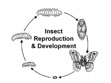
Holometabolous insects have immature forms
(larvae) that are very different from adults.
Larvae are "feeding machines", adapted mostly for
consuming food and growing in size. They become
larger at each molt but do not acquire any
adult-like characteristics. When fully grown,
larvae molt to an immobile pupal stage and
undergo a complete transformation. Larval
organs and appendages are broken down (digested
internally) and replaced with new adult
structures that grow from imaginal discs,
clusters of undifferentiated (embryonic) tissue
that form during embryogenesis but remain dormant
throughout the larval instars. The adult stage,
which usually bears wings, is mainly adapted for
dispersal and reproduction.
25
Larval Forms
Appearance Larval type Common Name
Description Example Campodeiform
Crawler flattened body with long legs
Neuroptera
usually w/
filaments on the end Trichoptera
of the abdomen Dytiscidae
Carabiform Crawler similar to
above, but legs are Chrysomelidae
shorter and filaments lacking Carabidae
Eruciform Caterpillar
cylindrical, well-formed head, Lepidoptera
thoracic legs, and abdominal
sawflies
prolegs
Scarabaeiform White grub C-shaped,
well-formed head. Scarabidae
and thoracic legs (no prolegs) weevils
26
Larval Forms
Appearance Larval type Common Name
Description Example Elateriform
Wireworm cylindrical, smooth, and
Elateridae tough skinned w/
short legs Tenebrionidae
Platyform None broad and
flat w/ legs short Syrphid fly or absent
blister beetle
Vermiform Maggot
cylindrical and elongate Diptera
lacks legs
Hymenoptera
Siphonaptera
27
Pupa
Appearance Pupal type Common Name
Description Example Obtect
Chrysalis Developing appendages held
Lepidoptera
tightly against
the body by a
shell-like
casing. Often found
enclosed
within a silken cocoon
Exarate None All developing
appendages free Coleoptera
and visible externally
Neuroptera Coarctate
Puparium Body encased within the hard
Diptera
exoskeleton of
the next-to-last
larval
instar
Recommended
