
Kein Folientitel - PowerPoint PPT Presentation
1 / 1
Title:
Kein Folientitel
Description:
Introduction. Persistent developmental stuttering (PDS) frequent ... right hemispheric (pre)motor ... 15 male subjects with PDS (mean age 26.7 years) ... – PowerPoint PPT presentation
Number of Views:49
Avg rating:3.0/5.0
Title: Kein Folientitel
1
Normal interhemispheric inhibition in persistent
developmental stuttering Martin Sommer, Kathrin
Knappmeyer, Evke Jane Hunter, Alexander Wolff
von Gudenberg, Nicole Spindler, Walter
Paulus Department of Clinical
Neurophysiology, University of Goettingen,
Germany, and Institut der Kasseler
Stottertherapie, Bad Emstal, Germany
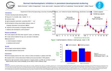
2.0
Control subjects
- Introduction
- Persistent developmental stuttering (PDS)
- frequent (1 of adults male female 4 1)
- unknown origin
- right hemispheric (pre)motor overactivity (PET)1,
2 but - left hemispheric timing abnormalities between
areas of - language preparation and execution (MEG)3
- impaired communication of language-related
areas4 - Impaired inter-hemispheric communication?
1.8
Stuttering subjects
1.6
Test pulse Left hemisphere
Test pulse Right hemisphere
1.4
MEP amplitude normalized to unconditioned (test)
MEP amplitudes
1.2
1.0
0.8
0.6
- Material and Methods
- 15 male subjects with PDS (mean age 26.7 years),
no cluttering. - 13 male controls matched for age, sex, and years
of education. - Measurement of
- Interhemispheric inhibition5
- Ipsilateral silent period6
0.4
Test
2
5
6
8
10
20
50
80
Test
2
5
6
8
10
20
50
80
Interstimulus interval ms
Figure 1 Interhemispheric inhibition in both
groups, mean /- SE.
35
Stuttering subjects
Control subjects
- Results
- Unchanged interhemispheric inhibition
- Unchanged ipsilateral silent period
30
25
20
Silent period ms
Figure 2 Ipsilateral silent period in both
groups, mean /- SE.
- Conclusions
- Normal interplay between the motor cortices of
either hemisphere in patients with PDS. - Consistent with a normal intracortical inhibition
previously described in PDS patients. - Abnormal right (pre)motor activity observed in
imaging studies on PDS are not likely to reflect
altered primary motor cortex excitability, but
are likely to have a different origin.
15
10
5
0
Left Hemisphere
Right Hemisphere
References 1. Fox P.T. et al., Nature 382, 158
(1996). 2. Braun A.R. et al., Brain 120, 761
(1997). 3. Salmelin R et al., Brain 123, 1184
(2000). 4. Sommer et al., Lancet 360, 380-83
(2002) 5. Interhemispheric inhibition measured
using a two-coil technique with the conditioning
pulse over the hand area of one and the test
pulse over the hand area of the other hemisphere
while the hand muscles were at rest. test pulse
adjusted to yield amplitudes of about 1.0 mV in
the abductor digiti minimi muscle, conditioning
pulse intensity adjusted to yield amplitudes of
about 1.5 mV. Interstimulus intervals of 2, 5, 6,
8, 10, 20, 50, and 80 ms studied 10 times each,
and unconditioned test stimuli 20 times.
Conditioned MEP amplitudes normalized to
unconditioned ones. Repeated-measures ANOVA with
interstimulus interval and side as within-
group and group as between-group factors. 6.
Ipsilateral silent period studied using a
one-coil technique over the hand area of one
motor cortex during voluntary activation of the
ipsilateral abductor digiti minimi muscle.
Stimulus intensity adjusted to yield MEP
amplitudes of about 2 mV contralaterally.
Duration of the induced ipsilateral silent period
obtained from 30 rectified trials was measured
and compared between groups using unpaired,
two-tailed t-test. Each side was investigated
separately, and the data were analyzed using a
repeated-measures ANOVA with side as within-
group and group as between-group factors.
Recommended
CrystalGraphics Presentations


