
Breast Imaging: Radiologic Pathologic Correlation - PowerPoint PPT Presentation
1 / 32
Title:
Breast Imaging: Radiologic Pathologic Correlation
Description:
Breast Imaging: Radiologic Pathologic Correlation – PowerPoint PPT presentation
Number of Views:641
Avg rating:3.0/5.0
Title: Breast Imaging: Radiologic Pathologic Correlation
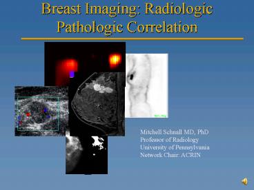
1
Breast Imaging Radiologic Pathologic Correlation
Mitchell Schnall MD, PhD Professor of
Radiology University of Pennsylvania Network
Chair ACRIN
2
Rad / Path Correlation
- Careful collaboration between radiology and
pathology - Cooperation of surgeon
- Method to index lesions/findings
3
Image analysis Turning an image into data
- User extracted qualitative features
- User extracted quantitative features
- Semi automated
- Automated
Feature 1 Feature 2 Feature 3 . . .
4
Abnormal enhancement descriptors
- focus
- mass
- non-mass
E. Morris, MSKCC
5
Invasive Ductal Carcinoma Irregular/Spiculated
6
FOCAL MASS ROUND/IRREGULAR/Rim Enhancing
7
Ductal Enhancement DCIS
8
Associated DCIS
9
(No Transcript)
10
(No Transcript)
11
Fibroadenoma Internal Septations
12
Fibroadenoma
13
Fibroadenoma
Post-Contrast
T2
Central component is glandular, bright T2, and
enhances Peripheral component is hyalinized, low
T2 and does not enhance
14
(No Transcript)
15
(No Transcript)
16
(No Transcript)
17
(No Transcript)
18
Multivariate model for Focal Mass
Model ROC Area Forward model 0.873 (0.871 to
0.875) Backward model 0.880 (0.877 to 0.884).
19
Image meta-dataStandard Structure and Data
Elements
Patient level data
Image Exam level data
Image Finding level data
Time
20
Modality 1 Time 1
Modality 2 Time 1
M1 Finding 2 Lesion 2
M1 Finding 1 Lesion 1
M2 Finding 2 Lesion 3
- M2 Finding 1
- Lesion 1
- Size
- Shape
- Location
- Intensity
- etc
Modality 1 Time 2
Path Lab Micro Proteomics
21
PET association with Receptor Status
22
(No Transcript)
23
(No Transcript)
24
Triple Neg
Non Triple Neg
25
Triple Neg
Non Triple Neg
26
Multimodaliy triple negative phenotype
- No spiculations on mammography
- No calcs on mammography
- Heterogeneous enhancement on MRI
- Well circumscribed on ultrasound
- Rim enhancement on MRI
- Elevated SUV on PET
27
Not Triple Negative Cancer
Triple Negative (Basal) Cancer
28
Not Triple Negative Cancer
Triple Negative (Basal) Cancer
29
Efficacy of MRI and Mammography for Breast-Cancer
Screening in Women with a Familial or Genetic
Predisposition
Kriege et al, NEJM 351, 2004 p.427
- 1909 women, 50 breast cancers detected, 45
Cancers detected by screening, 32 seen by MRI, 18
detected by mammography. (ROC analysis Plt0.5) - 13 cancers missed by MRI (8 seen on mammography)
30
Ca
No Ca
31
Interval cancer presentation
11/07
5/07
3/08
32
Collaborators
- Emily Conant
- Susan Weinstein
- Susan Orel
- Abass Alavi
- Mark Rosen
- Sara Englander
- Larry Dougherty
- Hee Kwon Song
- Joel Karp
- Sandy Seghal
Andrew Maidment et al John Tomaszewski Mike
Feldman Ray Boston Peter Moate Brian
Czernicki Susan Domchek Angie DeMichele Marcia
Boraas Sandy Schwartz
Recommended
CrystalGraphics Presentations


