
YFG cDNA - PowerPoint PPT Presentation
1 / 34
Title:
YFG cDNA
Description:
... transfectants of BHK cells (BHK = baby hamster kidney) Works: ... Mouse and hamster cell lines used for commercial ... data available for Chinese hamster ... – PowerPoint PPT presentation
Number of Views:131
Avg rating:3.0/5.0
Title: YFG cDNA
1
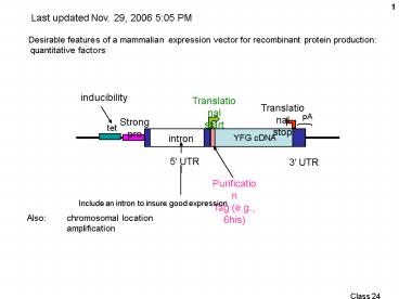
Last updated Nov. 29, 2006 505 PM
Desirable features of a mammalian expression
vector for recombinant protein production
quantitative factors
Translational start
Translational stop
Strong pro
YFG cDNA
intron
5 UTR
3 UTR
Include an intron to insure good expression
Also chromosomal location amplification
2
Desirable features of a mammalian expression
vector for recombinant protein production
qualitative factors
Altered posttranslational modifications
glycosylation Altered protein properties
improvements Creation of novel proteins
3
Penta- saccharide common core
Diantennary With bisecting GlcNAc With
fucosylated core
Triantennary (also tetra-antennary)
All shown here, N-linked (to amide N of Asn in
N-X-S or N-X-T)
Substantial in size
Carbohydrates attached to loops or near termini
Fucose
Also O-linked, to ser or thr (hydroxyl on side
chain)
4
Figure 7.28. Examples of O-linked
oligosaccharides O-linked oligosaccharides
usually consist of only a few carbohydrate
residues, which are added one sugar at a time.
5
Carbohydrate structure specific for Cell
type Physiological state No. of sites depends on
3-D structure of protein Structure at that site
depends on the site !
Example transferrin from different cell types
Cerebrospinal fluid (made in
brain) diantennary asialo agalacto fucosyla
ted bisecting GlcNAc Blood (made in
liver) diantennary NAcNeu (sialated sialic
acid) afucosylated
Sialic acid structure see next graphic
6
neuraminic acid one of the sialic acids
both terms are used, confusedly
NAcNeu
Carboxyl (acid)
Glycerol moiety
mannose
Acetylated amino group
deoxy
7
Glycosylation pattern affects signaling,
for Delivery to the right cell receptor for
activity Clearance rate
Microheterogeneity Lots of isoforms typically
present
Glycosylation does not seem to represent a
bottleneck in high-producing cells 0.1 mg/l ?
(amplify) ? 200 mg/l same pattern
Insect cells (Baculovirus, high level transient
expression) Too simple a pattern compared to
human
Mouse and hamster cells similar to
human Hamster less heterogeneity
8
Genetic engineering possibilities for
glycosylation Modify or enhance
activity E.g. Better binding to a
receptor More specific binding Different binding
Also Antigenicity reduce it Clearance rate
lower it Decrease microheterogeneity (for
clinical application) pure defined product
9
- Modifying glycosylation
- Add or subtract sites to your favorite protein
(cis) - 1a. Subtract sites Easy, change N or S or T to A
by site-directed mutagenesis - 1b. Add sites Not so easy.
- Consensus N-X-S does not work, e.g.
- requires the insertion of a 12 aa region
encompassing a real N-glycosylation site (6
suffices for O-linked) - Place on an end or on a loop (must know
proteins structure) - Works
- Change the general glycosylation phenotype of the
host cell (trans) - 2a. Clone cDNA for a glycosyl transferase from
one cell type or organism and trrasnfect it into
another cell type. - 2b. Lectin-resistant mutants exhibit different
deficiencies in glycosyl transferases (Pam
Stanley)
10
- Modifying glycosylation
- Add or subtract sites to your favorite protein
(cis) - Change the general glycosylation phenotype of the
host cell (trans)
2. Clone enzyme genesGlycosyl transferases,
mostlyAlso some synthetases (e.g., NAcNeu) Can
be complexe.g., 7 different fucosyl
transferases (FTs),with different (overlapping)
substrate specificities Simpler example
Construction and characterization of stably
transfected BHK-21 cells with human-type
sialylation characteristic. Cytotechnology 30
1725, 1999.Peter Schlenke1, Eckart Grabenhorst1,
Manfred Nimtz2 Harald S. Conradt1 Hamster
cells do only 2,3 sialylation. Humans do 2,6 as
well, via a 2,6 sialyl transferase
(ST) ExperimentOver-express cloned human 2,6
ST, along with a substrate protein.producing
permanent transfectants of BHK cells (BHK baby
hamster kidney) Works Get both types of
structures now, substantially (although not
exactly the same ratio as in human cells).
11
Isolate mutant mammalian cell lines deficient in
specific glycosylation enzymes
Stanley Isolation of multiply mutated
glycosylation mutants by selecting for lectin
resistance Lectins carbohydrate-binding
proteins Plant lectins used mostly here (but
occur widely) Sequential selections, push - pull
on resistance, sensitivity Resistance enzyme
deficiency ? failure to add the sugar need for
lectin binding Increased sensitivity failure
to add a sugar produces greater exposure of
underlying sugars in a transferase-negative
mutant ? better binding to the exposed
sugar Showed power of selection Showed
usefulness of complementation analysis via cell
hybridization Hybrid selection All lec-R
mutants were WGA (wheat germ agglutinin)
resistant (various degrees) pro- Tester
parent was single lec-R GAT- (req. glycine,
adenine and thymidine) Select in medium lacking
pro, GAT, and with /- WGA Complementing
hybrids will have regained sensitivity to
WGA Mutants in the same gene will remain WGA
resistant (non-complementation) Could now be
used as a tabla rasa for introducing a series of
enzymes to build custom tailored
glyco-conjugates. Complicated though (order of
addition, location in the Golgi, etc. )
Potential increased or deceased targeting to
carbohydrate-sensitive receptors (e.g., liver
asialoglycoprotein receptor) clearance
rate Review Annual Review of Genetics. 18
525-552 (December 1984) Glycosylation Mutants of
Animal Cells P Stanley
Pam Stanley
12
Umana, P., Jean-Mairet, J., Moudry, R., Amstutz,
H., and Bailey, J.E. 1999. Engineered glycoforms
of an antineuroblastoma IgG1 with optimized
antibody-dependent cellular cytotoxic activity.
Nat Biotechnol 17 176-180.
Target here (bisecting NAcG)
(NAcG N-acetyl-glucosamine here)
Presence of the bisecting NAcG enhances binding
of T-cell receptor to the Fc region of
antibodies. Binding is needed for ADCC. Mouse
and hamster cell lines used for commercial
production lack the glycosyltransferase needed
for bisecting NAcG addition A rat myeloma cell
line does produce MAb with the bisecting
NAcG. Hypothesis Expression of the rat enzyme
in a CHO cell line will add a bisecting NacG to
the anti-neuroblastoma MAb produced by these
cells. The modified MAb will be a better
mediator of ADCC. Experiment Clone the cDNA
for this enzyme from the rat line and transfer it
to CHO cells, driven by an inducible tet
promoter. Check sugar structure of MAb and ADCC
efficiency of the MAb.
13
ADCC assay
ADCC correlates with bisected complex content
Tet induction of GnTIII
No induction of GnTIII
14
Result ADCC efficiency followed proportion of
oligosaccharide with bisected sugar Bisecting
sugar15 ? 45 ADCC 25 ? 50
Missing Zero bisection control CHO cells are
supposed to LACK GnTIII Westerns show 0 rat
GnTIII at 2000 ug/ml tetracycline Yet
backgrounds are high. OK for ADCC, but Mass Spec
data .
Extensions?
Try untransfected CHO? Westerns lying? ( ug.ml tet ? death too much enzyme?)
Good example of enzyme engineering. Can still be
optimized.
Use a constitutive promoter, try different
version to find the best using ADCC as the
assay Check dependence on Ig production level.
15
Hypothesis Fucose interferes with binding of
the T-cell Fcgamma3 receptor to the Fc region of
an antibody molecule. Elimination of fucose from
produced MAbs will increase ADCC Create a mutant
CHO cells (starting with amplifiable dhfr- cells)
in which the fucose trasnferase genes have been
knocked out. All MAbs produced in these mutant
cells will be better at promoting ADCC
16
Double knock-out strategy for FUT8 an
alpha-1,6,fucosyl transferase
Little sequence data available for Chinese
hamster Isolate CHO cDNA using mouse sequence
data for primers Use CHO cDNA to isolate CHO
genomic fragments from a commercial lambda library
K.O. exon 1 translation start region
Homology regions
For hemizygote Select for G418
resistance, Screen by PCR for homologous recomb.
108 cells ? 45000 colonies? 40 false
recombinants (extension-duplications) 1 true
recombinant
Step 2 for homozygote, select for
Pur-resistance 1.6X10⁸?70,000 screened ? 10
double KO homozygotes.
Remove drug resis. genes by transient
transfection with Cre recombinase
Note 10s of thousands of PCRs performed to
screen for homologous recomb., using 96-well
plates
17
Double knockout evidence
After Cre treatment
Orginal KOd genes have a 1.5 kb
insertion (Southern blot)
mRNA has 200 nt deletion (RT-PCR
18
Use of a fluoresceinated lentil lectin (LCA) that
binds fucose oligosaccharides to demonstrate lack
of fucosylation in glycosylated proteins in the
FUT8 -/- cells
Control background fluorescence(FL-anti avidin)
FUT8 /
FUT8 /-
FUT8 -/-
19
Very laborious, but apparently a big
payoff. Better selection? Why not use the
fluorescent LCA to select for the FUT8 KOs along
with G418 resistance(double sequential
selection)?
/- heterozygotes?
AB overlaid
20
Rituxan (anti-CD20) produced in FUT -/- cells
does not contain fucose(HPLC analysis)
Digestion all the way to monosaccharides
Missing d - g
21
In ADCC, FUT8-/- anti-CD20 Rituxan
Binding to CD20 membranes FUT8-/- anti CD20
Ritxuan
Anti-CD20 from a partially FUT-deficient rat cell
line
Fc-Receptor protein binding assay
Rat line
FUT-/-s
Complement-mediated cell toxicity is the same
for FUT8-/- and Rituxan
Rituxan commercial product, 98 fucosylated
22
Second ( and further) generation recombinant DNA
therapeutic proteins More
improvements upon nature
Or Isolation of recombinant protein mutants
with altered binding properties
Why are they doing this? (TPA
tissue plasminogen activator) Problem Binding
of TPA to liver cells leads to clearance from the
bloodstream Want to avoid clearance in TPA
therapy (anti-thrombolytic, clot busting
protease) MAb387 to TPA blocks binding to
cultured hepatoma cells (liver-like cells) MAb387
decreases clearance rate. Goal Mutate the
cloned TPA gene. Mutate it in the MAb438-binding
region ? mutant TPA that Is hepatocyte
binding-negative (select for this ) Is still
protease (remains catalytically active) (screen
for this)
23
How could one do this? Select? Need to
characterize many mutant proteins, and find the
protein with the desired characteristic, and then
rescue the gene for that protein. Express in
mammalian cell transfectants But TPA is
secreted, so protein becomes divorced from the
DNA that coded it. My editorializing But
Coffino and Scharff had a technique for looking
at secretory variants (of myeloma cells)
Immunoprecipitate secreted proteins around
colonies grown in agar (Ig secretion,
precipitation by anti-mouse-Ig antibody)
24
Coffino and Scharff (Proc Natl Acad Sci U S A.
1971 Jan68(1)219-23.)
Alternative 1
Medium in agar
All in soft agar
Imagine Antibody in top layer MAb387 Colonies
CHO cell permanent co-transfectants of mutant
library TPA
Takes longer. Colonies may not make enough. But
you dont need a FACS ()
Precipitate -
Precipitate
25
Alternative 2 Phage display a way to link the
variant protein to its coding DNA Mutagenize the
gene as a fusion protein to a phage coat protein
and make a library in bacteriophage. The mutants
will be displayed on the surface of the phage and
can be panned for (or against).
DNA
DNA
DNA
Here, on would collect the members of the phage
library of mutant TPAsthat dont stick to
hepatoma cells, or to immobilized MAb328. But
lots of noise in a negative selection
non-stickers could be for many uninteresting
reasons (denatured, statistical, etc )
26
Rice et al. here used mammalian cells as the
carrier of the DNA and the cell surface as a
display site. Somewhat analgous to phage
display. Fusion protein to a membrane anchor
protein (peptide, really DAF decay
accelerating factor). What did they
do? Mutagenesis What region? 333 bp K1
(kringle-1), known to bind the MAb387, an
antibody that competes for hepatocyte binding
(so kringle-1 is likely to contain the hepatocyte
binding domain). How did they get it
mutated? Error-prone PCR How did they isolate
just the kringle 1 region? As a PCR
fragment. How did they get the mutagenized
fragment back in? Introduced restriction sites at
the ends, w/o affecting the protein coding.
27
What did they put the mutagenized fragment
into? TPA-DAF fusion protein DAF
decay-accelerating factor, anchored to
surface via phosphatidyl inositol joined to its
carboxyl terminal prevents cell lysis by
complement Last 37 AAs of DAF suffices How
did they get the TPA-DAF gene into into
cells? Electroporation How many copies per cell.
And why is that important? One, by
electroporation at low DNA concentration. In a
transient transfection! Binding is dominant.
Lack of binding is recessive. How did they
select cells making MAb387-non-binding TPA? FACS,
sorting cells with low fluorescence from the
MAb387, But high fluoresence from MAb372, that
binds to the protease domain.
28
(No Transcript)
29
How did they recover the plasmid carrying the
mutant TPA gene from the selected cells? Hirt
extraction Like a plasmid prep, high MW DNA
allowed to get entangled and form a
clot. Centrifuge. Chromosomal DNA ? soft
pellet plasmid DNA circles stay in supernatant.
Then re-transfect, re-sort in FACS. After 2
sorting rounds, test individual E. coli clones
60 are binding-negative. Clone mutated regions
into regular TPA gene for testing Assayed
hepatoma cell binding. How?Label WT TPA with
fluorescein (FITC) (conjugate chemically) Mix
with hepatoma cells and analyze on a flow
cytometer (FACS w/o the sorter part). See
specific and non-specific binding. Subtract
non-specific binding the amount not competed by
excess un-labeled wt TPA.
30
Rice et al Fig.2
FACS analysis
MAb to protease domain
enriched
Low kringle-1 reactivity
MAb to kringle-1 domain
31
AND
FACS selection can also work for an internal
protein (Urlaub et al. 1985)
by absence of
Est. freq. of DHFR- mutants 10-4
Still only 1 in 10 were mutants.
32
Rice et al Fig. 4
WT
Compete. So still bind.
33
(No Transcript)
34
Amino acid changes define the domains responsible
for hepatocyte binding and protease activity.
Amino acid changes in the interior are probably
folding mutants (lose all activities)
Recommended
CrystalGraphics Presentations


