
Continuation of Lecture 14 - PowerPoint PPT Presentation
1 / 20
Title:
Continuation of Lecture 14
Description:
Provides the pathway for tracts between higher and lower brain centers ... Three paired fiber tracts that connect the cerebellum to the brain stem ... – PowerPoint PPT presentation
Number of Views:105
Avg rating:3.0/5.0
Title: Continuation of Lecture 14
1
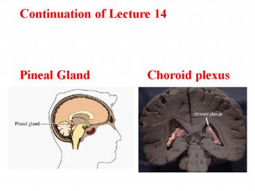
Continuation of Lecture 14 Pineal
Gland Choroid plexus
2
Epithalamus
3
Brain Stem
Consists of three regions Similar to spinal
cord but contains embedded nuclei Controls
necessary for survival Provides the
pathway for tracts between higher and lower brain
centers Associated with 10 of the 12 pairs of
cranial nerves
4
Brain Stem
5
Midbrain
Located between the and the Midbrain
structures include two bulging structures
that contain descending pyramidal motor
tracts hollow tube that connects the third
and fourth ventricles Various nuclei
6
Midbrain Nuclei
Nuclei that control cranial nerves III ( ) and
IV ( ) four domelike protrusions of the
dorsal midbrain visual reflex centers
auditory relay centers functionally
linked to basal nuclei largest nucleus of
the reticular formation red nuclei are relay
nuclei for some descending motor pathways
7
Midbrain Nuclei
8
Pons
Bulging brainstem region between the and
the Forms part of the anterior wall of the
fourth ventricle Fibers of the pons Connect
higher brain centers and the spinal cord Relay
impulses between the motor cortex and the
cerebellum Origin of cranial nerves V ( ), VI
( ), and VII ( ) Contains nuclei of the
reticular formation
9
Pons
10
Medulla Oblongata
Most inferior part of the Along with the pons,
forms the ventral wall of the fourth
ventricle Contains a choroid plexus on the
ventral wall of the fourth ventricle Pyramids
two longitudinal ridges formed by corticospinal
tracts Decussation of the pyramids crossover
points of the corticospinal tracts
11
Cerebellum
Located dorsal to the Protrudes under the
occipital lobes of the cerebrum Makes up 11 of
the brains mass Provides precise timing and
appropriate patterns of skeletal muscle
contraction Cerebellar activity occurs
subconsciously
12
Anatomy of the Cerebellum
Two bilaterally symmetrical hemispheres connected
medially by the vermis transversely oriented
gyri Each hemisphere has three lobes anterior,
posterior, and flocculonodular gray matter
cortex, internal white matter, scattered
nuclei distinctive treelike pattern of the
cerebellar white matter
13
Cerebellar Peduncles
Three paired fiber tracts that connect the
cerebellum to the brain stem All fibers in the
cerebellum are Superior peduncles connect the
to the Middle peduncles connect the to
the Inferior peduncles connect the to the
14
Protection of the Brain
The brain is protected by Harmful substances are
shielded from the brain by the Meninges Three
connective tissue membranes lie external to the
CNS mater, mater, and mater Functions
Cover and protect the CNS Protect blood vessels
and enclose venous sinuses Contain cerebrospinal
fluid (CSF) Form partitions within the skull
15
Meninges
16
Blood-Brain Barrier
Protective mechanism that helps maintain a
stable environment for the brain Bloodborne
substances are separated from neurons
by Continuous endothelium of Relatively
thick basal lamina Bulbous feet of
17
Spinal Cord
CNS tissue is enclosed within the vertebral
column from the to Provides two-way
communication to and from the brain Protected
by space between the vertebrae and the
dural sheath (dura mater) filled with fat and a
network of veins
18
Spinal Cord
terminal portion of the spinal cord
fibrous extension of the pia mater anchors the
spinal cord to the coccyx delicate shelves
of pia mater attach the spinal cord to the
vertebrae
19
Spinal Cord
- 31 pairs attach to the cord by paired roots
- sites where nerves serving the upper and
lower limbs emerge - collection of nerve roots at the inferior
end of the vertebral canal
20
Spinal Cord
- Gray matter consists of soma, unmyelinated
processes, and neuroglia - connects masses of gray matter encloses
central canal - Posterior (dorsal) horns interneurons
- Anterior (ventral) horns interneurons and
somatic motor neurons - Lateral horns contain sympathetic nerve fibers
White Matter Fibers run in three directions
Divided into three funiculi (columns)
posterior, lateral, and anterior Each funiculus
contains several fiber tracks Fiber tract names
reveal their origin and destination Fiber tracts
are composed of axons with similar functions
Recommended
CrystalGraphics Presentations


