
I' Basics of Electron Microscopy - PowerPoint PPT Presentation
1 / 20
Title:
I' Basics of Electron Microscopy
Description:
Tungsten crystal (field emission) 100x brighter ... X. Helical Reconstruction. planar 2D lattice. Cylindrical (helical) 2D lattice ... – PowerPoint PPT presentation
Number of Views:479
Avg rating:5.0/5.0
Title: I' Basics of Electron Microscopy
1
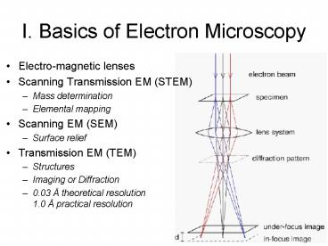
I. Basics of Electron Microscopy
- Electro-magnetic lenses
- Scanning Transmission EM (STEM)
- Mass determination
- Elemental mapping
- Scanning EM (SEM)
- Surface relief
- Transmission EM (TEM)
- Structures
- Imaging or Diffraction
- 0.03 Å theoretical resolution1.0 Å practical
resolution
2
II. Electron Source
- Tungsten filament (thermionic)
- Tungsten crystal (field emission) 100x brighter
- Electrons Accelerated to defined energy behave as
a wave. - ??kV (100 1000 kV with 200kV-gt 0.025 Å)
3
III. Lenses and deflection coils
- Lenses focus beam and form images
- Coils control beam direction
4
IV. Column Design
- Inverted light microscope
- Gun
- Condenser lens(es)
- Objective lens
- Projection lens (ocular)
- Apertures
5
V. Detectors
- Film 3.5 x 4 cm, small grain size
- CCD fast readout, good dynamic range, poor
resolution - Imaging plates excellent dynamic range, high cost
6
VI. Specimen Preparation
- Vacuum Conductivity Contrast Radiation
Damage - SEM gold coat
- STEM freeze-dry, carbon coat
- TEM
- plastic section positive stain
- Protein suspension/crystal
- Negative stain
- Frozen-hydrated
7
VII. Structure Determination
- Tissue
- tomography
- Crystals
- crystallography
- Isolated complexes
- single particle analysis
8
VIII. 3D reconstruction
- Projection theorem
- Tilt Reconstruction
9
IX. 2D crystallography
- Fourier Transform composed of 2D lattice lines
- Individual Projections sample lattice lines
10
IX. 2D Crystallography (cont)
- Electron Diffraction amplitudes
- Images phases
- Max tilt /- 60º missing cone
11
X. Helical Reconstruction
planar 2D lattice
Cylindrical (helical) 2D lattice
12
X. Helical reconstruction (cont)
13
X. Helical reconstruction (cont)
14
X1. Electron tomography
EM control
Data collection
60
coordination
-60
CCD control
15
(No Transcript)
16
3D-reconstruction by back projection
medical imaging - CAT scan, MRI
mathematical foundation
Horiz proj
17
(No Transcript)
18
Reconstructed Image Segmentation
Plakoglobin plakophilin
cadherin
membrane
desmoplakin
Sample Thickness 50 nm CCD DATA
0.931 nm/pixel Theoretical resolution
2.1 nm Practical Resolution 4-5nm or better
Intermediate Filament
19
(No Transcript)
20
(No Transcript)
Recommended
CrystalGraphics Presentations


