
Garry W' K' Ho, M'D' - PowerPoint PPT Presentation
1 / 52
Title: Garry W' K' Ho, M'D'
1
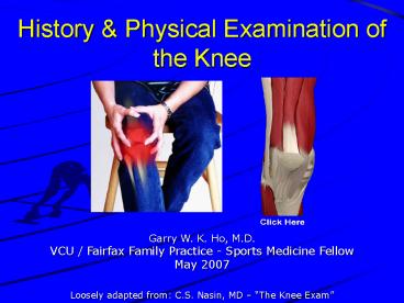
History Physical Examination of the Knee
Garry W. K. Ho, M.D. VCU / Fairfax Family
Practice - Sports Medicine Fellow May 2007
Loosely adapted from C.S. Nasin, MD The Knee
Exam
2
Objectives
- Review pertinent clinical anatomy of the knee
- Review differential diagnosis of knee complaints
- Review clinical history and physical examination
of the knee - Common knee injuries findings
Shaun Livingston
3
Anatomy - Osteology
4
Anatomy - Osteology
- Femur
- Medial lateral
- Condyles
- Epicondyles
- Trochlear groove
- Intercondylar notch
- Patella
- Superior pole (base)
- Inferior pole (apex)
- Medial lateral facets
- Tibia
- Medial lateral
- Condyles
- Gerdys tubercle
- Pes anserine area
- Tibial tuberosity
- Tibial plateau
- Tibial spines
- Fibula
- Head
- Neck
5
Anatomy Major Ligaments Tendons
- Quadriceps tendon
- Patellar tendon
- Medial lateral patellar retinaculum
- Medial collateral ligament (MCL)
- Lateral collateral ligament (LCL)
- Iliotibial tract (IT band)
- Anterior cruciate ligament (ACL)
- Posterior cruciate ligament (PCL)
6
Anatomy Menisci of the Knee
- Medial meniscus
- Lateral meniscus
- Meniscal ligaments
- Functions of the menisci
- Meniscal zones
- White-white
- Red-white
- Red-red
7
Anatomy Popliteal Area
- Popliteal fossa
- Popliteal vessels
- Tibial nerve
- Common peroneal nerve
- Gastrocnemius
- Medial lateral heads
- Hamstrings
- Semitendinosus
- Semimembranosus
- Biceps femoris
- Popliteal (Baker) cysts
8
Anatomy Bursae about the Knee
9
Differential Diagnosis
- Osteoarthritis / Chondromalacia
- Inflammatory arthritis
- Meniscal tears
- Avascular necrosis
- Fractures
- Supracondylar femur
- Patellar
- Tibial plateau
- Osteochondral lesions
- Dislocations / subluxations
- Tibio-femoral
- Patello-femoral
- Patellofemoral Pain syndrome
- Plica syndrome
- Apophysitis
- Osgood-Schlatter
- Sinding-Larsen Johanssen
- Tendonosis
- Quadriceps
- Muscle and tendon strains
- Myotendinous contracture syndromes
- Iliotibial tract (band)
- Hamstring
- Bursitis
- Supra-, Pre-, Infra- patellar
- IT band, pes anserine
- Myositis ossificans / Heterotopic ossification
- Nerve entrapment
- Common peroneal
- Infrapatellar nerve
- Tibial nerve
- Vascular diseases
- Peripheral vascular disease
- Neuro-claudication
- Referred pain
- Intraartivular hip disease DDD
- Slipped Capital Femoral Epiphysis
- Radiculopathy
10
Clinical History
- Mechanism of Injury ? Able to continue play?
11
Clinical History
- Location of pain
- Onset Timing
- Acute vs. Chronic
- Traumatic vs. Overuse
- Characterize pain
- Night pain
- Morning stiffness
- Weakness
- Deformity
- Instability / Giving Way
- Locking / Clicking / Popping /
- Catching / Clunking
- Alleviating / Exacerbating Factors
- Previous treatments
- Sport Exercise / Training equipment habits
- Occupation
- History of prior injury
- Other symptoms (ROS)
12
Physical Examination
- Observation
- Undress waist ? down
- Shorts
- Palpation
- Active passive ROM
- Special tests
- Extensor mechanism, effusions, anterior knee
- ACL PCL
- MCL LCL
- Meniscal tests
- Contracture testing
13
Observation
- Undress waist ? down
- Gown, drape, or shorts
- Gait pattern
- Deformity
- Ecchymosis / Abrasion
- Erythema / Edema
- Joint Effusion (more later)
- Atrophy
- Limb length discrepancy
- Foot ankle morphology kinematics
14
Surface Anatomy Palpation - Anterior
15
Surface Anatomy Palpation - Medial
16
Surface Anatomy Palpation - Lateral
17
Surface Anatomy Palpation - Posterior
18
Range of Motion
- Passive
- Active ROM
- Flexion 135
- Extension 0 to -5
- Internal rotation 10
- External rotation 10
19
Intra-articular Effusions
- Large effusions
- Fairly obvious on inspection
- Moderate-large effusions
- Patellar ballottement
- Smaller effusions
- Milk effusion fluid
- Suprapatellar pouch ? distally to side ? tap
- Look feel for fluid wave
20
Extensor Mechanism Integrity
- Straight-leg Raise
- Quadriceps tendon
- Patella
- Patellar tendon
21
Anterior Knee Patellofemoral Joint
- Patellar instability
- Patellar Apprehension
- Lateral patellar displacement
- ? patient apprehension
- or guarding
- Patellar Glide
- Knee at 30 deg flexion
- Medial lateral patellar displacement
- Measured in quadrants
- Normal 1-2 quadrants
22
Anterior Knee Patellofemoral Joint
- Patellar instability
- Patellar tracking
- Patellofemoral pain
- Palpation of patellar facets
- Glide and lift patella medially laterally
- Palpate undersurface of patella for tenderness
23
Anterior Knee Patellofemoral Joint
- Patellofemoral pain
- Patellar Grind Test
- AKA Tether test
- Knee 10 deg flexion
- Glide patella distally, and firmly compress
patella against trochlear groove - Active quadriceps contraction ? pain
24
Anterior Stability - ACL Injury
- Symptoms
- Pain, audible pop
- Unable to RTP
- Swelling within hours
- Symptoms of instability
- Mechanism of Injury
- 80 - Non-contact
- Plant, deceleration, pivot on a planted foot
- Soccer
- Femur ? ER on fixed tibia
- Valgus load
- Basketball
- Hyperextension
- IR of tibia
- Contact mechanism
25
Anterior Stability - ACL Injury
- Anterior Drawer Test
- Knee flexed to 90 degrees
- Anteriorly translate tibia on femur
- Watch feel for amount of translation end
point - Lachman Test
- Knee flexed to 15-30 degrees
- Stabilize distal femur
- Anteriorly translate tibia on femur
- Watch feel for amount of translation end
point - Other Tests
- Pivot-Shift Test, etc
26
Posterior Stability - PCL Injury
- Symptoms
- Often vague
- Insecure feeling, possibly symptoms of
instability - Diffuse aching knee pain
- Difficulty climbing stairs
- Mechanism of Injury
- Dashboard injury
- Posteriorly directed force
- ? anterior aspect of
- flexed knee
- Falling onto flexed knee
- With foot plantar flexed
27
Posterior Stability - PCL Injury
- Posterior Drawer Test
- Knee flexed to 90 degrees
- Posteriorly translate tibia on femur
- Watch feel for amount of translation end
point - Sag Sign
- Knees flexed, quads relaxed
- ? compare both sides
- Look for tibial posterior sag relative to femur
- Quad-Active Test
- Knee flexed hamstrings fully relaxed
- Slide foot along table (quad active)
- Observe for anterior relocation
28
Valgus Stability - MCL Injury
- Symptoms
- /- medial swelling
- Medial pain TTP
- /- symptoms of instability
- /- lateral ecchymosis
- Mechanism of Injury
- Direct blow to lateral knee
- ? valgus stress
- Plant twist
- ? valgus stress
- Valgus stress to knee on planted leg)
- Unhappy Triad
29
Valgus Stability - MCL Injury
- Valgus Stress Testing
- Knee flexed to 30 degrees
- Relax ACL/PCL joint capsule
- Valgus stress applied to knee
- Look and feel for translation and endpoint
- Compare to uninjured side
- May repeat with knee in full extension
30
Varus Stability - LCL Injury
- Varus Stress Testing
- Same test as valgus stress testing
- Except applying a varus stress instead
- LCL, IT band, PLC are tested
31
Meniscal Injuries
- Symptoms
- Pain
- Medial
- Lateral
- Poorly localized
- Pain usually worse with squatting stairs
- Popping, catching, locking, or buckling
- Delayed effusion
- Unless peripheral tear
- Mechanism of Injury
- Non-contact cutting, deceleration, hyper-flexion
- Poorly landing from jump
- Medial gt Lateral
- Unhappy triad
32
Meniscal Injuries
- Joint line tenderness
- Medial or lateral
- Squat duck-walk
- No pain significant meniscal tear unlikely
- McMurray Wilson Tests
- Medial meniscus
- Flexed knee
- Apply valgus external rotation stress
- ? Extend knee
- Lateral meniscus
- Flexed knee
- Apply varus internal rotation stress
- ? Extend knee
- Positive painful click
33
Meniscal Injuries
- Apley meniscal compression distraction tests
34
Indications Ottawa Knee Rules
- Stiell et al. - Ann Emerg Med 1995
- Ottawa Knee Rules
- Age 55 or older
- Isolated tenderness at patella (no bony
tenderness of knee other than patella) - Tenderness at head of fibula
- Inability to flex knee to 90 degrees
- Patient unable to bear weight for four steps
immediately and in the emergency department or
office - Sensitivity 100 ? 97
- NPV 100
- Specificity 49 ? 27
35
Muscle Contracture Testing
- Musculotendinous tightness
- Predispose to injury
- Hamstring
- IT Band
- Hip flexors
36
Summary
- Know your anatomy and injury patterns
- Know your differential
- diagnoses
- You diagnose what you know
- Get a detailed history
- Stay organized
- Know why youre doing
- an exam
- Follow-up, follow-up, follow-up
37
Questions ?
38
Thanks !
39
Hamstring Contracture Testing
- Popliteal angle
- Hamstring contractures
- Patient supine
- Hip flexed to 90
- Passively extend knee
- ? until resistance encountered
- Normal is lt 40
40
Hip Flexion Contracture Testing
- Modified Thomas Test
- Patient sitting on edge or end of exam table
- Holding unaffected knee in flexion
- Affected hip and knee relaxed
- ? Patient instructed to freely / swiftly lie
back, holding unaffected knee in flexion - Positive test affected hip and knee pops up
into flexion - May be more sensitive
41
IT Band Contracture - Obers Test
- Lateral decubitus with testing side up, testing
knee flexed - Adduct and fully flex hip ? Abduct, externally
rotate, extend hip - Maintain this hip position
- Slowly release support against gravity from leg,
allowing gravity to take leg towards
- Positive test for IT band / TFL contracture
- Leg remains abducted or fails to return to
adducted position despite releasing leg - Positive test for gluteus medius contracture
- Test positive with hip in neutral flexion
42
Pivot Shift Test
- Test for rotational instability in ACL tears
- Knee in extension
- Secure lateral leg near knee
- Secure medial leg just distal to other hand
- Apply internal rotational gentle valgus force
to the extended knee - Then bring knee ? flexion
- Observe for clunk
- Reduction of the anterolaterally subluxed tibia
on femur by IT band - Usually at about 30 flexion
43
Posterolateral Corner
- IT Band
- Biceps femoris tendon
- LCL
- Posterolateral joint capsule
- Lateral meniscus
- Lateral coronary ligament
- Popliteus
- Arcuate ligament complex
- Arcuate ligament
- Popliteofibular ligament
- Fabellofibular ligament
44
Posterolateral Corner Injuries
- More significant injury
- Posterolateral instability
- Rotational instability
- Associated with other ligamentous injuries
- Atraumatic, chronic laxity vs. traumatic injury
- Very poor prognosis without prompt surgical
repair - Often missed ? need for high index of suspicion
- Subtle exam findings
- Hyperextension
- External Rotation Recurvatum Test
- Posterolateral Drawer
- Reverse Pivot Shift Test
- Dial Test
Easily Missed Diagnosis Ahead
45
Hyperextension External Rotation Recurvatum
Test
- Supine position
- Grasp great toes of both feet
- Lifts both LE off exam table
- Compare both knees
- Assess for increased knee
- Extension ? hyperextension or recurvatum
- External rotation of tibia
- Varus
- Positive ? severe ligament injury
- Combined PLC / or ACL and PCL injury
46
Posterolateral Drawer
- Similar to posterior drawer
- Knee flexed to 90
- Foot rotated 15 - 20
- Apply posterior external rotational stress
- Assess for posterolateral translation endpoint
of tibia on femur - Positive test ? presence and severity
47
Reverse Pivot Shift Test
- Basically the pivot shift done in reverse
- Knee flexed to 60- 70
- Secures foot / ankle
- Secure lateral leg near knee
- Apply external rotational force to the flexed
knee - Then bring knee towards full extension
- Observe for clunk
- Reduction of posterolateral subluxation of tibia
on femur by IT band
48
Dial Test (AKA Thigh-Foot Angle)
- Supine, seated, or prone position
- External rotation
- at 30 90 degrees
- Abnormal if
- gt10 degrees (absolute)
- gt5 degree side to
- side difference
- Compare to
- contralateral side
49
Review of Evidence ACL
(Jackson JL, et al.)
- Lachman Test Sens 87 Spec 93
- Anterior Drawer Sens 48 Spec 87
- Pivot Shift Test Sens 61 Spec 97
50
Review of Evidence - Meniscus
(Jackson JL, et al.)
- Joint Line Tenderness Sens 76 Spec 29
- McMurray Test Sens 52 Spec 97
51
Review of Evidence - General
(Ebell MH.)
52
Sample Management Algorithm
Recommended
CrystalGraphics Presentations


