
Cardiac Morphogenesis - PowerPoint PPT Presentation
1 / 42
Title: Cardiac Morphogenesis
1
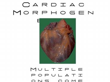
Cardiac Morphogenesis
- Multiple populations come together to make a heart
2
The mammalian heart is a two-sided pump that
provides oxygenated blood to the body and
unoxygenated blood to the lungs
- Your heart has four chambers
- The ventricles are the major pumping chambers
- The right side is the pulmonary side
(unoxygenated) - The left side is the systemic side(oxygenated)
- Note in the embryo, both sides are full of
oxygenated blood since oxygen comes from mom and
the lungs are underwater
3
Heart Development
- Brian Black (brian.black_at_ucsf.edu)
- Heart Development
- morphogenesis
- conduction
- valves
- epicardium
- second heart lineage
- cardiac neural crest
- tissue-tissue interactions reciprocal signaling
- Discussion
- Chang CP, Neilson JR, Bayle JH, Gestwicki JE, Kuo
A, Stankunas K, Graef IA, Crabtree GR. A field
of myocardial-endocardial NFAT signaling
underlies heart valve morphogenesis. Cell. 2004
Sep 3118(5)649-63.
4
Congenital cardiac anomalies are the most common
birth defects in the United States
- 40,000 babies are born with cardiac defects in
the United States each year (1125 live births) - Congenital heart defects are the leading cause of
birth defect-related deaths in the United States - Ventricular septal defects (VSD) and outflow
tract alignment and septation defects are among
the most severe forms of CHD
Sources March of Dimes, American Heart
Association
5
The heart begins as a crescent after gastrulation
and loops to form a four-chambered structure
6
A bilateral region of anterior lateral mesoderm
gives rise to the heart
7
A cardiac field is specified in the anterior
lateral mesoderm as cells ingress through the
streak at the time of gastrulation
8
Signals from the notochord/ventral neural
tube, ectoderm and endoderm promote
cardiac competence to a region of the anterior
lateral mesoderm
9
Wnt and BMP signals pattern the crescent
10
The heart comprises multiple lineages
pericardium
11
- The cardiac conduction system
12
The conduction system controls the heartbeat
The conduction system is derived from myocytes
that undergo trans-differentiation from a muscle
phenotype to a more electrically active phenotype
13
The conduction system is critical for a regular
heart beat
Purkinje fibers are critical to contraction and
to arrhythmia
co-expression of endothelin converting enzyme
(ECE-1) and endothelin in the embryonic heart is
sufficient to convert cardiomyocytes into
Purkinje fibers
ECE-1 expression in the heart is controlled by
hemodynamics induced by pressure and stretch
14
- Heart Valves
15
The heart has four valves, which control blood
flow and develop from endothelial cells that
receive a combination of signals, including BMP,
SHH, and others--these signals initiate EMT and
migration out of the endothelial layer
16
Valves form primarily from endothelial cells that
undergo EMT (epithelial to mesenchymal
transformation--often called endocardial to
mesenchymal transformation in this case--and
migrate into the jelly
17
Cardiac cushions will form the valves and are
responsible for separation of the outflow
vesselsCushions are composed primarily of
endothelial cells, but also consist of cardiac
neural crest cells in the outflow valves
The pulmonic valve is shown here
18
VEGF signaling functions via NFAT transcription
factors to control valve morphogenesis
Cell 118 532-534
19
The Epicardium
20
The septum transversum, or proepicardial organ,
resides just caudal to the heart and will give
rise to the epicardium and coronary vessels as
well as the liver, for which it is better known
heart
Septum transversum
21
The septum transversum gives rise to the
coronaries and the epicardium
The epicardium responds to retinoic acid from the
myocardium by producing TGF-ß, which is required
for myocardial growth during development
22
- The Second Heart Lineage
23
Birds
Mammals
Two mesodermal progenitor populations contribute
to the heart
24
The second heart lineage contributes to the
outflow tract and right ventricle, and numerous
genes and enhancers have activity restricted to
these regions of the heart, which suggested a
distinct developmental domain
25
Numerous, common congenital anomalies occur in
the secondary heart field or where the primary
and secondary fields are joined... Double outlet
right ventricle (DORV) Persistent truncus
arteriosus (PTA) Some ventricular septal defects
How are these distinct cell populations integrated
at the time of cardiac looping?
26
(No Transcript)
27
(No Transcript)
28
Tetralogy of Fallot
29
The second heart lineage contributes to the OFT,
RV, and some LV but not many other parts of the
heart
30
Outflow tract myocardium and endothelium are
AHF-derived but the smooth muscle appears to be
neural crest-derived
31
- The Cardiac Neural Crest
32
Neural crest cells are multipotent progenitor
cells
Craniofacial mesenchyme
Outflow tract smooth muscle
33
Neural Crest
Neural crest tissue found ONLY IN
VERTEBRATES Forms melanocytes, much of the
craniofacial skeleton, peripheral glial cells,
peripheral autonomic neurons, and chromaffin
cells of the adrenal gland, AND IS ESSENTIAL FOR
OUTFLOW TRACT AND AORTIC ARCH ARTERY DEVELOPMENT
34
Fate map of the neural crest along the A-P axis
The adopted fates are due to signals encountered
during migration
Le Douarin, N. M. et al. Development
20041314637-4650
35
Cranial neural crest cells migrate along distinct
pathways and give rise to craniofacial mesenchyme
and contribute to the aortic arches, outflow
tract, and the cardiac cushions
Trigeminal stream-jaws, zygome, palate, majority
of face and skull
Hyoid stream
Post otic stream--outflow tract and aortic arches
36
Neural crest cells contribute to the aortic arch
arteries AND provide essential signals for their
remodeling
37
(No Transcript)
38
Neural Crest and second heart lineage each
contribute to the outflow tract Neural crest
required for septation Second heart lineage
required for alignment But remember, these
populations are together in the arches and are
talking to each other
39
Defects in Neural crest migration contribute to
multiple anomalies in humans
40
Numerous, common congenital anomalies occur in
the secondary heart field or where the primary
and secondary fields are joined
- Double outlet right ventricle (DORV) and other
outlet alignment defects - Persistent truncus arteriosus (PTA)
- Some ventricular septal defects
The formation of the outflow tract, ventricular
septum, and valves requires complex interplay
between the secondary heart field and the neural
crest
Bmp4 cond. KO
Overriding Aorta VSD, Eric Jaehnig
Persistent Truncus Arteriosus VSD, Dave McCulley
41
Multiple progenitor populations are involved in
cardiac morphogenesis
42
Reciprocal signaling is critical to heart
development and to vertebrate organogenesis in
general
- Neural crest and the second heart lineage
communicate with each other to set outflow tract
elongation, alignment, and septation - Myocardium signal to the endocardium, which
stimulates EMT and valve formation - Myocardium signals to epicardium, which in turn
signals back to the myocardium to promote
myocardial growth
Recommended
CrystalGraphics Presentations


