
New Dimensions In Medical Imaging - PowerPoint PPT Presentation
1 / 32
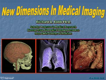
Title: New Dimensions In Medical Imaging
1
New Dimensions In Medical Imaging
Richard A. Robb Ph.D.
Scheller Professor in Medical Research Director
Mayo Biomedical Imaging Resource Mayo Clinic
College of Medicine
2
The Evolving Dimensions Of Medical Imaging
- Old 2D x-ray, nuclear
- Recent 3D CT, MRI, PET, etc.
- New 4D dynamic CT, MRI, US
- Horizon 5D 4D function
3
Imaging Over The Space Dimension (Scale)
Macro Micro
Pulmonary parenchyma (millimeters)
Pulmonary epithelial cells (microns)
4
Modern Medical Imaging
- Faster (dynamic)
- Farther (3D anatomy)
- Functional (physiology)
- Fusion (anatomy function)
5
Modern Biomedical Imaging
Scanners Computers Users
Apply
Acquire
Process
6
3D/4D Volume Image Acquisition Systems
CT
MRI
PET
PET/SPECT
7
Advanced Image Processing Used In Medical Image
Applications
- Enhancement
- Rendering
- Segmentation
- Modeling
- Fusion
8
Image Enhancement
Enhancement of pulmonary structures
Before
After
Restoration of lung epithelial cells
Before
Correction of MR field bias
After
After
Before
9
Volume Image Rendering
Raw 2D Image Stack
3D Image Volume Rendering
10
Realistic Tissue Rendering
11
Volume Image Segmentation
12
Volume Image Modeling
Patient-Specific Geometrically,
Texturally Functionally Accurate Computer
Models of Anatomy, Physiology, Pathology
13
Volume Image Registration/Fusion
14
5D Heart Fusion From Patient Data
15
Image Guided Clinical Applications
- Brain Surgery For Epilepsy
- Orthopedic Surgery Planning
- Virtual Colonoscopy
- Cardiac Arrhythmia Ablation
- Conjoined Twins Separation
16
Image-Guided Neuro-Surgery (For Treatment of
Epilepsy)
Subtraction Inter-ictal Spect CO-registered to
MRI - SISCOM
3D SPECT functional images taken before and after
seizure, subtracted, and registered to 3D MRI
anatomic image for precise localization of
seizure focus to plan and guide resection of
diseased tissues
Target tissue can be displayed in 2D or 3D
17
Orthopedic Surgery Planning
3D CT scans and xrays can be used to precisely
plan the size, shape and location of surgical
implants, including hip or spine prostheses
18
Quantitative Virtual Colonoscopy
Polyp
1.8cm
Micro-vessels in Polyp
19
Cardiac Arrhythmia - Atrial Fibrillation
- Increasing Prevalence
- 1 incidence for 60 yo, 10 for 80 yo
- 2.3 M patients in 2001, 5.8 M by 2050
- 45 patients with class IV failure have Afib
- Unbridled Progression
- 50 recurrent, 25 to chronic over 5 yr
- Accompanying Mortality/Stroke
- 50 mortality/stroke over 10 yrs age 55-75
- Skyrocketing Costs
- Hospitalization more than doubling
- How to Treat ??
- Drugs, Pacemakers, Surgery, Ablation
20
Treating Atrial FibrillationWith Image-Guided
Catheter Ablation
The goal is to accurately find see the target,
precisely navigate to it, effectively ablate, and
monitor the result.
21
5D Image-Guided Cardiac AblationHardware-Software
platform that integrates and dynamically
displays cardiac electrophysiology mapped onto 3D
cardiac anatomy in real time to rapidly and
accurately guide catheter ablation of arrhythmias
(Funded by NIH/NIBIB EB-002834)
(US Patent 6,556,695 ER Patent 224,
621 2003)
22
Real-time Image-Guided Ablation Process
23
Separation of Conjoined Twins - 1991
24
Separation of Conjoined Twins - 1993
Before
Surgical Planning
3D rendering of torsos
Volume CT Scan
Calculation of opening areas
After
Planning optimal bisection
25
Separation of Conjoined Twins - 2006
26
Image Processing and Analysis
Selective 3D Anatomy Segmentation
Medical Illustrations
Relative liver perfusion by each twin
Life-size physical model
27
Separation of Conjoined Twins - 2006
28
Success!!
29
Impact of Modern Medical Imaging On Clinical
Practice
- Improved outcomes and yield
- Reduced risk and morbidity
- Less procedure time, faster recovery
- Higher throughput and less cost
30
Elements For Continuing Progress
It is not always be easy, but we make progress
with
ideas
Past
Future
tools
and teamwork modern
and future science is multi- inter-disciplinary
i.e., you cant do it alone!
The only easy day was yesterday! (US Navy Seal
Motto)
31
Poised For The Future
- Real-time Minimally Invasive Technology
- Synchronous Diagnosis Treatment
- New Imaging Modalities
- Molecular Biochemical Imaging
- Intelligent Instruments Robots
- In vivo seek-treat-monitor nano-bots
- Seamless Fusion Of Structure Function
- Real-time integration, Organ ? Molecule
32
The Fantastic Future
From Star Trek IV Courtesy Paramount
Pictures
Recommended
CrystalGraphics Presentations


