Chapter 5 Protein Function - PowerPoint PPT Presentation
1 / 27
Title:
Chapter 5 Protein Function
Description:
16.7 kDa globular, 153 residue O2-binding protein. Highly conserved in many organisms ... Steric considerations affect binding of other molecules to heme (CO, CN ... – PowerPoint PPT presentation
Number of Views:128
Avg rating:3.0/5.0
Title: Chapter 5 Protein Function
1
Chapter 5 Protein Function
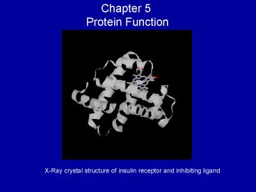
X-Ray crystal structure of insulin receptor and
inhibiting ligand
2
Protein-ligand interactions case studyMyoglobin
(Mb)
- ? 16.7 kDa globular, 153 residue O2-binding
protein - Highly conserved in many organisms
- Found in high quantity in muscle
- Coordinated to Fe2
- Contains the heme prosthetic group
- Reversibly binds O2 for delivery to cells
3
Myoglobin Structure and FunctionThe heme group
RED Porphyrin group
Hexa-coordinated iron ion (Fe2)
- Iron is in the Fe (II) oxidation state, which
binds reversibly to molecular O2. - Q. What prevents oxidation to Fe (III)?
- Electron-donating character of N atoms in
porphyrin ring and - Group is buried deep in interior of protein
4
Fe is coordinated to 4 N atoms of heme, N of His
93, leaving one site for O binding
Fe2
5
O2 Binding
- Binding requires access of O2 to the deeply
buried heme - Protein breathing allows transient openings to
interior of protein - Steric considerations affect binding of other
molecules to heme (CO, CN-)
6
Secondary and tertiary structure
7
Protein-ligand equilibrium
Definition ? ? fraction of binding sites on
protein occupied by ligand Kd Ligand
concentration at which ½ of sites are
occupied (Half-saturation) NOTE Low Kd means
high protein affinity for ligand
8
Myoglobin (P)-O2 (L) Binding
- For gases, use PRESSURE, so expression for ?
changes to
where P50 is the partial pressure of O2 at half
saturation
- P50 of Mb 2.8 torr
- P(O2) 100 torr (arterial) and 30 torr (venous)
- Thus, Mb remains bound to O2 over wide range of P
9
Hemoglobin (Hb)
- 4-subunit, 65 kDa protein, (4? structure)
consisting of myoglobin-like units - Hb is an allosteric protein (binding effects
subsequent binding) - 2 ? subunits 2 ? subunits
- Interactions between subunits at interface are
hydrophobic, H-bonds, electrostatic - 1 Hb binds 4 O2
- P nL ? PLn
- Hb-O2 dissociation curve is not described by
hyperbola
10
Mb vs. Hb
Mb binds O2 at pressures at which Hb releases it!
11
Slope n, degree of Cooperativity n lt 1
Negative cooperativity n gt 1 Positive
cooperativity n 1 Non-cooperative
12
Hb-O2 (OxyHB) Binding Mechanism
- T (tense) state Predominates in low O2
environment (deoxyHB) - Upon O2 binding, T ? R (relaxed) state (oxyHb)
- R state is stabilized by ligand (O2, CO, CN-)
binding T state by salt bridges - Conformation change is result of alterations in
subunit interfacial contacts and interactions - Changes in heme structure (pucker vs. planar)
account for cooperativity and sigmoidal
dissociation curve
13
Transport of CO2 and HThe Bohr Effect
(Christian, Niels daddy)
- In periphery, PCO2 is high pH is low
- Low pH decreases Hbs affinity for O2
- CO2 H2O ? H HCO3- (catalyzed by carbonic
anhydrase) - Hb(O2)nHx O2 ? Hb(O2)n1 xH
- So, H and Hb-O2 affinity are inversely
proportional - CO2 binding also decreases Hb affinity for O2
- Protonation stabilizes T-state by affecting
subunit interactions
14
BPG
- Why does stripped Hb have a higher O2 affinity
than Hb in whole blood? - Negative charges interact via salt bridges to
basic (positively charges) amino acids in Hb - BPG decreases Hb-O2 affinity, allowing
dissociation at lower pO2 - FHb (fetal Hb) has lower affinity for BPG than
does AHb (adult) this means HIGHER O2 affinity - High altitude acclimation involves production of
BPG
15
Immunology
- Highly specific interactions between protein (T
cell receptors and antibodies) and antigens (any
agent that elicits a cellular immune response - Humoral system (fluid)
- Antibodies and immunoglobulins (Ig)
- Produced by B lymphocytes
- Bind and bacteria, viruses, foreign compounds
- Cellular system
- T lymphocytes (T cells cytotoxic T cells)
- Cells contain T cell receptors proteins on cell
surface which recognize and bind antigens - Helper T cells produce cytokines (e.g.,
interleukins) which stimulate T cell
proliferation
16
How Not to Kill Yourself
- MHC proteins Distinguish between pathogens and
self - MHC Classes I and II
- Present protein fragments on cell surface
- Foreign peptides are recognized as such by T cell
receptors and targeted for destruction - T cells against self proteins are not allowed to
mature (in thymus) - Class II MHC proteins utilize helper T cells to
stimulate response - HIV targets these cells, significantly disturbing
immune response
17
(No Transcript)
18
(No Transcript)
19
Fab Antigen binding fragment Fc Base of Y
20
Types of Antibodies
- IgA, IgD, IgE, IgG, IgM
- IgM is earliest present in immune response
- IgG is major in secondary response (memory B
cells) - Leukocytes are activated, which destroy invaders
by phagocytosis
21
(No Transcript)
22
Antibody-antigen interactions are the basis for
many analytical techniques
- Polyclonal antibodies Many B lymphocytes raised
against 1 antigen - Bind different parts of same antigen
- Monoclonal antibidies Raised in identical B
cells against 1 antigen - Whole population binds the same site on antigen
- Both can be used, e.g., for affinity
chromatography - ELISA (Enzyme-linked immunoabsorbant assay)
- Protein of interest linked to subphase
- Solution containing antibody is introduced binds
(TIGHTLY) to protein - Secondary antibody (raised against first
antibody attached to enzyme that catalyzes
color-producing reaction) is introduced - Immunoblot assay
- Proteins are transferred from PAGE to membrane
and washed with primary and secondary antibodies - Linked enzyme catalyzes signal producing reaction
- See movie!
23
(No Transcript)
24
Actin-Myosin SystemEnergy-requiring motility
- Based on protein-protein interactions
- Myosin (6-subunit protein thick filiment)
contains several helices and ATP-binding domain - Actin monomers aggregate to form thin filament
- Actin walks along myosin, resulting in muscle
contraction and relaxation
25
(Actin)
(Myosin)
26
(No Transcript)
27
Muscle Contraction Regulation
- 2 proteins, troponin and tropomyosin, control
actin-myosin muscle contraction - Tropomyosin (in complex with troponin) binds
actin, inhibiting actin-myosin interaction - Ca, released as response to stimulus, binds
troponin, causing conformational change, allowing
actin-myosin interactions (contraction)

