
1' The Respiratory Tract - PowerPoint PPT Presentation
1 / 28
Title:
1' The Respiratory Tract
Description:
external intercostal muscles. accessory muscles. respiratory muscles during expiration: ... internal intercostal muscles. abdominal walls. Lung pressures ... – PowerPoint PPT presentation
Number of Views:87
Avg rating:3.0/5.0
Title: 1' The Respiratory Tract
1
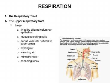
RESPIRATION
- 1. The Respiratory Tract
- A. The upper respiratory tract
- Nose
- lined by ciliated columnar epithelium
- mucus secreting cells
- dense vascular network in submucosa
- filtering air
- warming air
- humidifying air
- sneezing reflex
2
- Pharynx
- Epiglottis
- Larynx
- vocal cords
- glottis
- B. The lower respiratory tract
- Trachea
- Bronchi
- Bronchioles
- Respiratory bronchioles
- Alveolar ducts and alveoli
3
- Conducting zone
- to warm and humidify the air
- to distribute the gas
- to serve as part of body defense system
- Respiratory zone
4
- Respiratory tract defense system
- Mucocilliary transport system mucus escalator
- Cough reflex
- Macrophages
5
- 2. The Lung Mechanics
- A. Lung pressures and ventilation
- The thorax and respiratory muscles
- thoracic cage ribs (12), sternum, diaphragm
- pleural space
- respiratory muscles during inspiration
- diaphragm
- external intercostal muscles
- accessory muscles
- respiratory muscles during expiration
- Diaphragm
- internal intercostal muscles
- abdominal walls
6
- Lung pressures
- Air flows because of pressure gradients
- pleural pressure (Ppl)
- alveolar pressure (PA)
- Pressure changes during respiratory cycle
- pneumothorax
7
- Lung volumes and capacities
- Spirometry
- tidal volume (VT)
- inspiratory reserve volume (IRV)
- expiratory reserve volume (ERV)
- residual volume (RV)
- inspiratory capacity (IC)
- functional residual capacity (FRC)
- vital capacity (VC)
- total lung capacity (TLC)
- forced vital capacity (FVC)
8
- FEV1 timed forced expiratory volume in one
second - FEV1/FVC 80 more useful for detecting
obstructive vs restrictive lung diseases
9
- Minute respiratory volume (V, minute ventilation)
- V VT f (respiratory rate)
- Dead space volume (VD)
- Alveolar ventilation (VA) VA (VT - VD) f
10
- B. Mechanical Properties of the lung
- Lung Distensibility
- Pressure-volume curve
- Compliance (CL DV/DP)
- Pulmonary surfactant
- surface tension
- Laplace Law P 2T/r
- atelectasis
11
- Work of breathing
- W force X distance
- Factors that affect the amount of work
- lung compliance
- surface tension
- airway resistance
- R ? L ? /r4
- diameter of the airways
- Bronchoconstriction histamine
- Broncodilation CO2, EP (?2 receptors)
12
- 3. Pulmonary Circulation
- A. Vascular pressure and blood flow
- Pulmonary circulation is a low-pressure system
- pulmonary arterial systemic pressure 25 mmHg
- pulmonary arterial diastolic pressure 10 mmHg
- mean pulmonary arterial pressure 15 mmHg
- effect of the special gravity of blood on
distribution of blood flow in the lung - poor perfusion in the upper lung (functional dead
space volume)
13
- Hypoxic vasoconstriction balances blood flow with
ventilation - regional hypoxia/hypoxemia
- hypoxic vasoconstriction - a mechanism that
balances the perfusion of blood with the
availability of regional ventilation - effect of hypoxic vasoconstriction at the high
altitude - Exercise recruits capillaries and decreases
transit time
14
- 4. Gas Uptake and Transport
- A. Gases diffuse through respiratory membrane
- Daltons law PB PO2 PCO2 PN2 PH2O PHe
- barometric pressure PB at the sea level 760
mmHg - partial pressures
- PO2 PB X F O2 760 X 0.21 160 mmHg
- vapor pressure of water
- PO2 in alveolar gas and venous blood 100/40 mmHg
15
Gas exchangealveoli and cells
16
- Factors that affect the rate of gas diffusion
through the respiratory membrane - thickness of respiratory membrane
(alveolar-capillary membrane) normally 0.1 - 0.5
µm - pulmonary edema
- fibrosis of the lung
- surface area of the respiratory membrane 70 m2
in the normal adult - emphysema (dissolution of alveolar walls)
- diffusion coefficient
- solubility in water
- molecular weight
- carbon dioxide diffuses 20 times as rapidly as
oxygen - pressure difference across the respiratory
membrane
17
Respiratory membrane
18
- Pulmonary pathologies
19
- B. Transport of oxygen
- Transport of oxygen in the dissolved state
- only 2 of oxygen transported in the dissolved
state in the water of the plasma and cells - Transport of oxygen by hemoglobin
- 98 oxygen is carried to the tissues by
reversible combination with hemoglobin - oxygen carrying capacity 20 ml/100ml blood
- oxygen saturation percent O2 saturation O2
content/O2 capacity x 100 - oxyhemoglobin dissociation curve
- factors that affect the oxyhemoglobin curve
20
(No Transcript)
21
Oxygen-hemoglobin dissociation
"2,3-DPG and oxygen/Hb binding"
- Factors that affect the oxyhemoglobin curve
22
- Factors contributing to the total oxygen content
of arterial blood
23
(No Transcript)
24
- D. Control of Breathing
- Neural mechanisms
- Medullary respiratory centers
- inspiratory neurons set the rhythm
- expiratory neurons
- receive synaptic inputs from the cortex and pons
- effects of pulmonary stretch receptors
(proprioreceptors) - failure of the respiratory center
- by physical damages (concussions, cerebral edema)
- by overdose of chemical substances (barbiturate,
anesthetics)
25
- Reflex control of ventilation
- Chemoreceptors monitor blood gases and pH
- Control centers in the brain stem regulate
activity to respiratory muscles
26
- Chemical mechanisms
- chemoreceptors
- central chemoreceptors (in the medulla) monitor
only H in CSF - peripheral chemoreceptors (aortic bodies and
carotid bodies) - control of the alveolar ventilation by the
arterial CO2 - control of the alveolar ventilation by the
arterial H exclusively by peripheral
chemoreceptors - control of the alveolar ventilation by the
hypoxia relatively insensitive to hypoxia
27
Central chemoreceptor
Carotid body oxygen sensor
28
Chemoreceptor reflex
Recommended
CrystalGraphics Presentations


